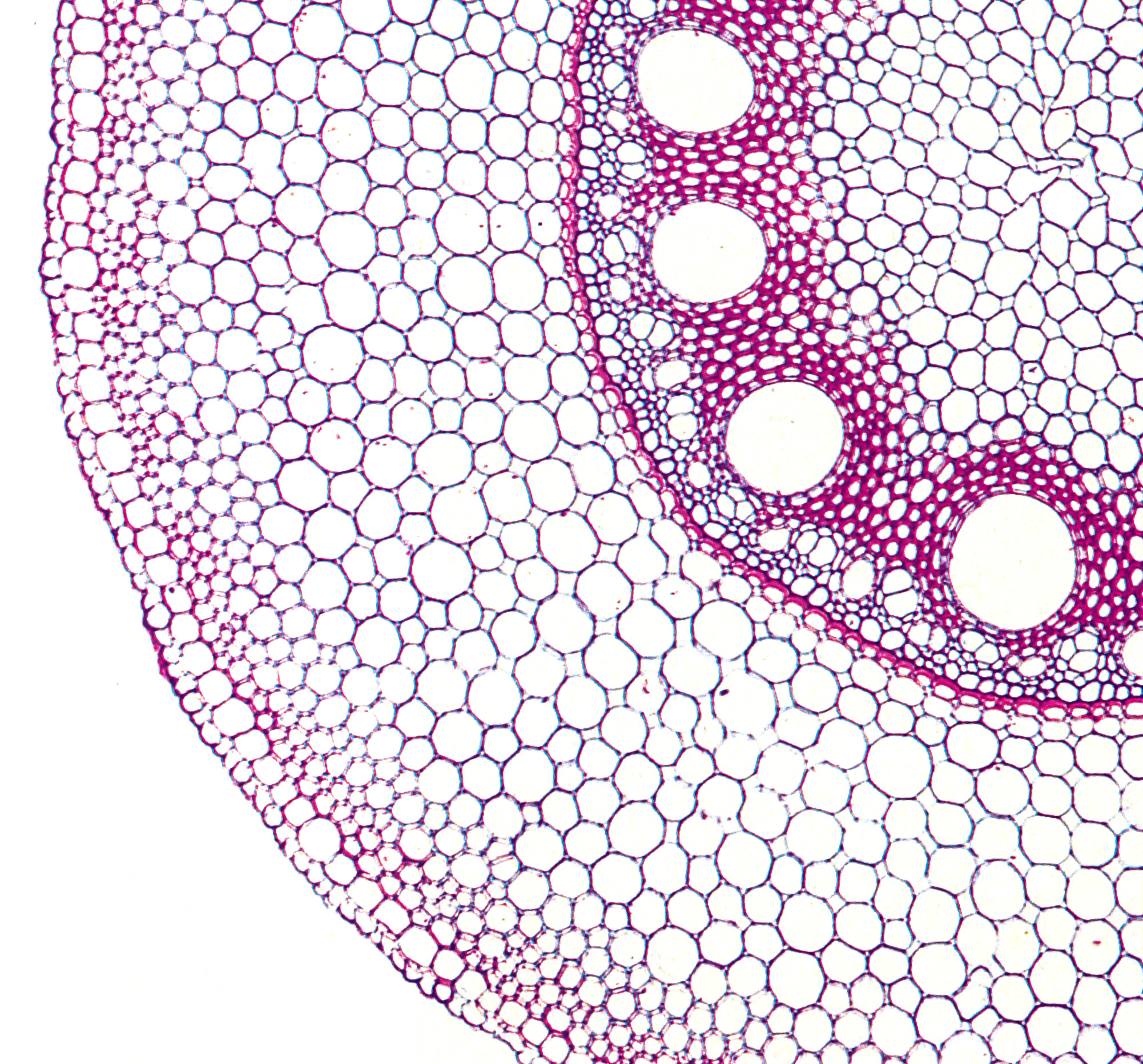
Figure 3.46. Transverse section of a mature maize root, showing many layers of cortical cells outside a distinctive suberised endodermis bounding the stele. Late metaxylem vessels with large diameters are the dominant feature of the stele. Note a hypodermal layer underlying the epidermis. Pink Toluidine blue staining characterises suberised cells. (Photograph courtesy A.W.R. Robards)