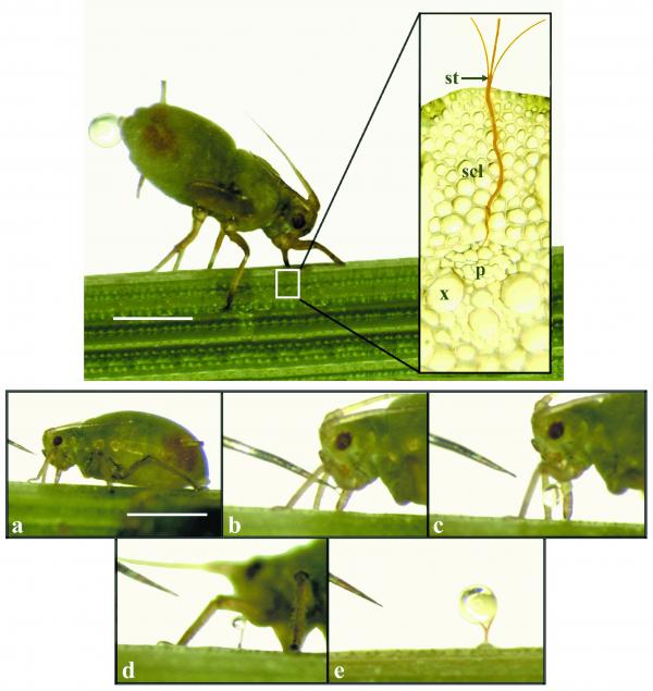
Figure 5.8 Aphids can be used to collect phloem sap. Top photograph: a feeding aphid with its stylet embedded in a sieve tube (see insert); scl, sclerenchyma; st, stylet; x, xylem; p, phloem. Note the drop of ‘honeydew’ being excreted from the aphid’s body. Plates (a) to (e) show a sequence of stylet cutting with an RF microcautery unit at about 3-5 s intervals (a to d) followed by a two-minute interval (d to e) which allowed exudate to accumulate. The stylet has just been cut in (b); droplets of hemolymph (aphid origin) are visible in (b) and (c); once the aphid moves to one side the first exudate appears (d), and within minutes a droplet (e) is available for microanalysis. Scale bars: top = 1 mm, bottom = 1.5 mm (Courtesy D. Fischer)
