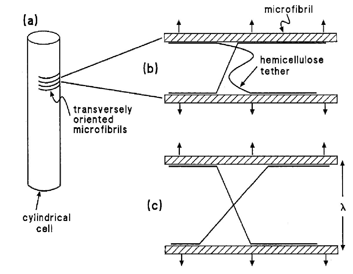
Figure 7.21 Schematic portrayal of the structure of an expanding cell wall. (a) A cylindrical cell, showing the transverse arrangement of cellulose microfibrils. (b) A pair of adjacent microfibrils connected by hemicellulose molecules (tethers) that are attached by hydrogen bonds; the arrows denote the force generated by turgor that is moving the microfibrils apart. One of the tethers, the straight one, is load-bearing; the other (wavy) is not. (c) The same microfibrils as in (b), whose separation in response to the applied force has resulted in a previously loose tether becoming load-bearing. (JB Passioura and SC Fry, Aust J Plant Physiol 19: 565-576, 1992)