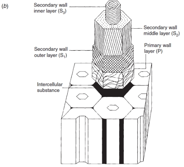
Figure 7.23 Schematic representation of the wall of a fully grown cell, showing the cellulose microfibril orientation in the primary (P) and secondary (S1, S2, S3) wall layers of a xylem fibre cell or a tracheid. (AB Wardrop and DE Bland, In ‘Biochemistry of Wood’ Pergamon Press, 1959)