Nicolas L. Taylor1,2 and A. Harvey Millar1
1ARC Centre of Excellence in Plant Energy Biology and 2School of Chemistry and Biochemisty, The University of Western Australia.
During photosynthesis the carbon assimilated is either retained in the chloroplast as starch or converted to sucrose and directed for export to sites of growth. Starch is degraded by a series of enzymes in the chloroplast, with sucrose degradation mainly occurring in the cytosol and both leading to glycolysis and the oxidative pentose pathway which produce respiratory substrates. These carbon rich compounds are prime sources of respiratory substrates in plants, although other carbohydrates such as fructans and sugar alcohols are also used.
During respiration, metabolites are oxidised and the electrons released are transferred through a series of electron carriers to O2. Water and CO2 are formed and energy is captured as ATP which is harnessed to drive a vast array of cellular reactions.
In comparison to sucrose and starch, the contribution of proteins and lipids as sources of respiratory substrates in most plant tissues is minor; exceptions to this generalisation are the storage tissues of seeds such as castor bean and soybean, in which amino acids and lipids may provide respiratory substrates, and during the processes of senescence in plant tissues where protein and lipid degradation increases.
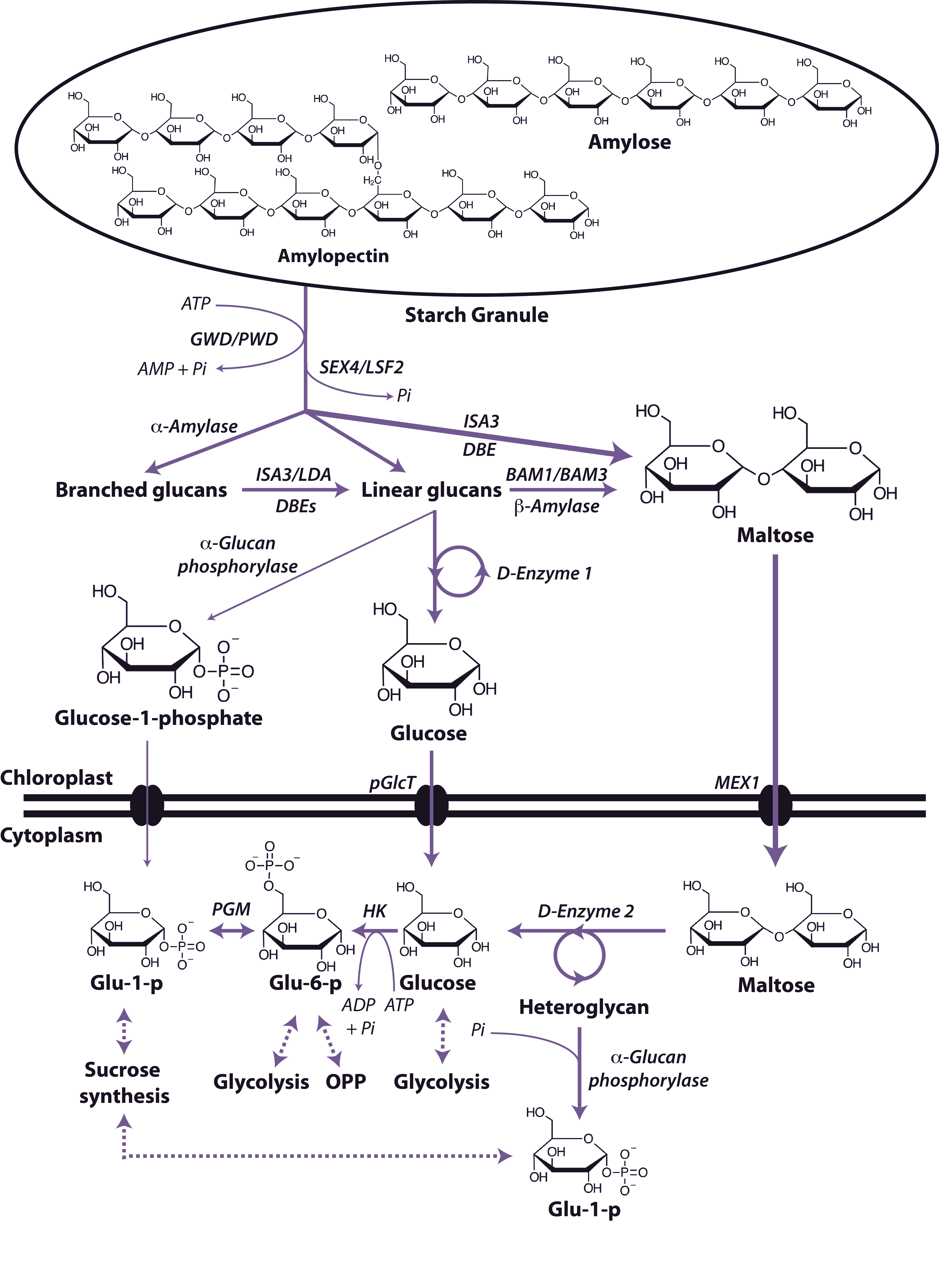
Starch is the principal storage carbohydrate in plants and this carbon reserve plays a number of important roles in plants. It is composed of two polymers of glucose, amylose and amylopectin and is stored in the plastid (chloroplast in leaves, amyloplasts in non-photosynthetic tissues) as insoluble, semi-crystalline granules. Starch is accumulated during rapid growth in the day and is almost completely degraded at night to mostly glucose and maltose, which is exported from the chloroplast and metabolised in the cytosol (Figure 2.19). Starch degradation is initiated by the addition of phosphate groups at the C6-position and C3-position of individual glucosyl residues that act to disrupt the packing of the glucans at the granule surface. These phosphate additions are catalysed by two enzymes, glucan water dikinase (GWD) and phosphoglucan water dikinase (PWD) respectively. The hydrolysis of the resulting glucan and phosphoglucan chains is carried out by a suite of enzymes including the phosphoglucan phosphatases (SEX4/LSF2), β-amylases (BAM1/BAM3), debranching enzymes (DBE; ISA3/LDA), α-amylase (AMY3), α-glucan phosphorylase and the disproportionating enzyme 1 (D-enzyme 1; an α-1,4-glucanotransferase).). The resulting maltose and glucose are exported to the cytosol by the glucose transporter (pGlcT) and maltose transporter (MEX1) and glucose-1-phosphate is thought to be exported by a similar but yet unknown mechanism. Once in the cytosol the maltose and glucose are converted to substrates for either sucrose synthesis, glycolysis or the oxidative pentose phosphate pathway by a number of enzymes including the disproportionating enzyme 2 (D-enzyme 2; an α-1,4-glucanotransferase), α-glucan phosphorylase, hexokinase and phosphoglucomutase.
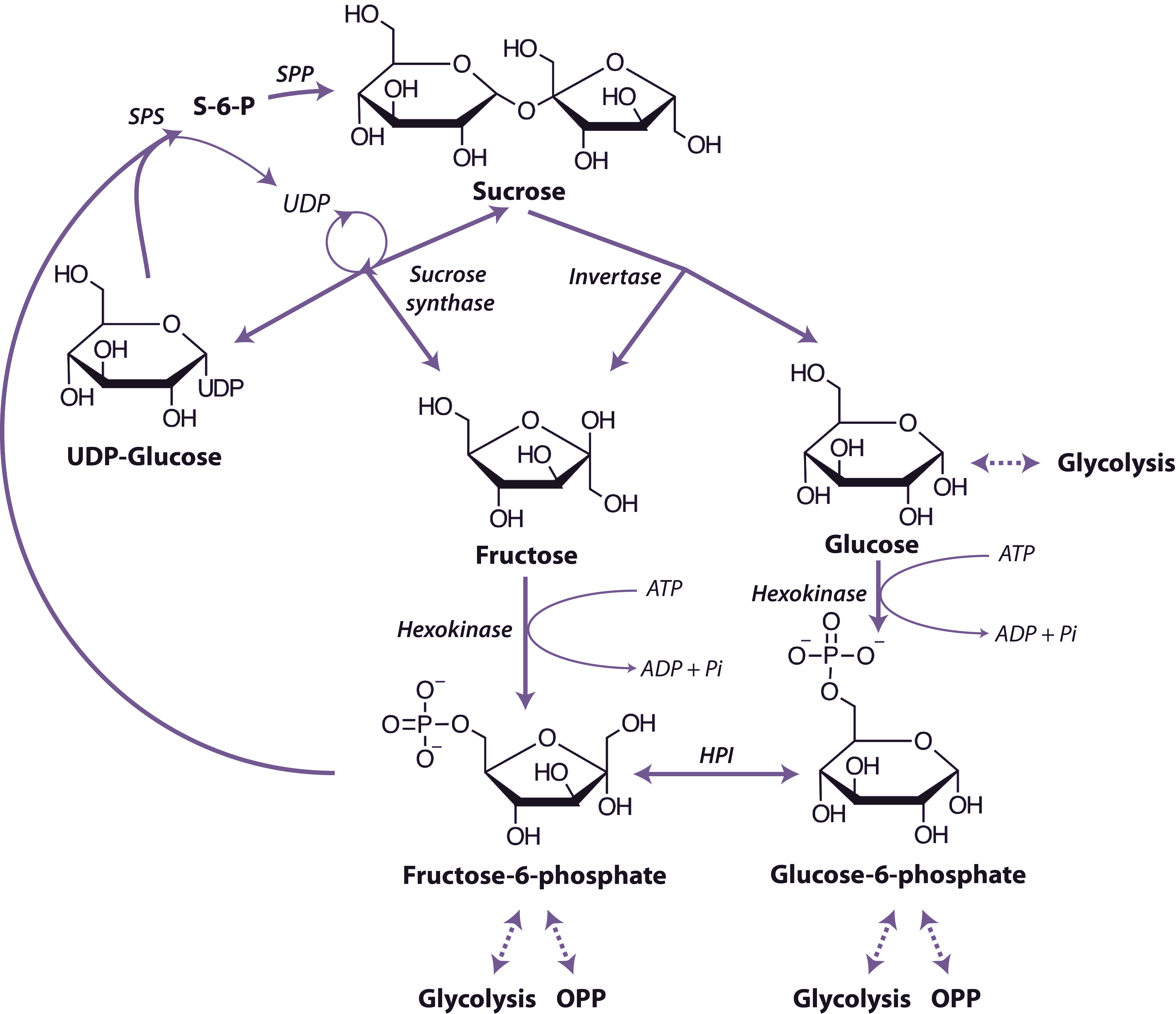
Sucrose is the world’s most abundant disaccharide, it is only produced by photosynthetic organisms and serves a role as a transportable carbohydrate and sometimes as a storage compound. The reactions in plant tissues leading to degradation of sucrose to hexose monophosphates are outlined in Figure 2.20.
The first step is cleavage of the glycosidic bond by either invertase (Equation 2.1) or sucrose synthase (Equation 2.2).
\[\text{Sucrose + H}_2\text{O} \rightarrow \text{D-Glucose + D-Fructose}\tag{2.1}\]
\[\text{Sucrose + UDP} \rightarrow \text{UDP-Glucose + D-Fructose}\tag{2.2}\]
Plant tissues contain distinct invertases located in the vacuole, cell wall (acid invertases) cytosol, mitochondria, nucleus, and cholorplast (neutral/alkaline invertases) which hydrolyse sucrose to glucose and fructose in an irreversible reaction. The invertases are differentially regulated by a number of mechanisms including pH to allow them to function in cell expansion, supply of carbon skeletons and energy metabolism. Multiple isoforms of sucrose synthase are located in the cytosol or cytosolic membranes that catalyse a thermodynamically reversible reaction, but this reaction probably acts only to breakdown sucrose in vivo. Their activity is developmentally regulated and they have functions in the supply of activated glucose for starch and cellulose biosynthesis. While both invertase and sucrose synthase can both breakdown sucrose, research using knockouts of multiple isoforms of both enzymes has shown that sucrose synthase is not required for normal growth in Arabidopsis, whereas invertase is indispensable. However this does not rule out the requirement of sucrose synthase in certain tissues of crop plant tubers, seeds and fruits where it has been shown to be crucial. Glucose and fructose are metabolised further by phosphorylation to the corresponding hexose-6-P by hexokinase. Hexokinase in plant tissues is associated with the outer surface of mitochondria.
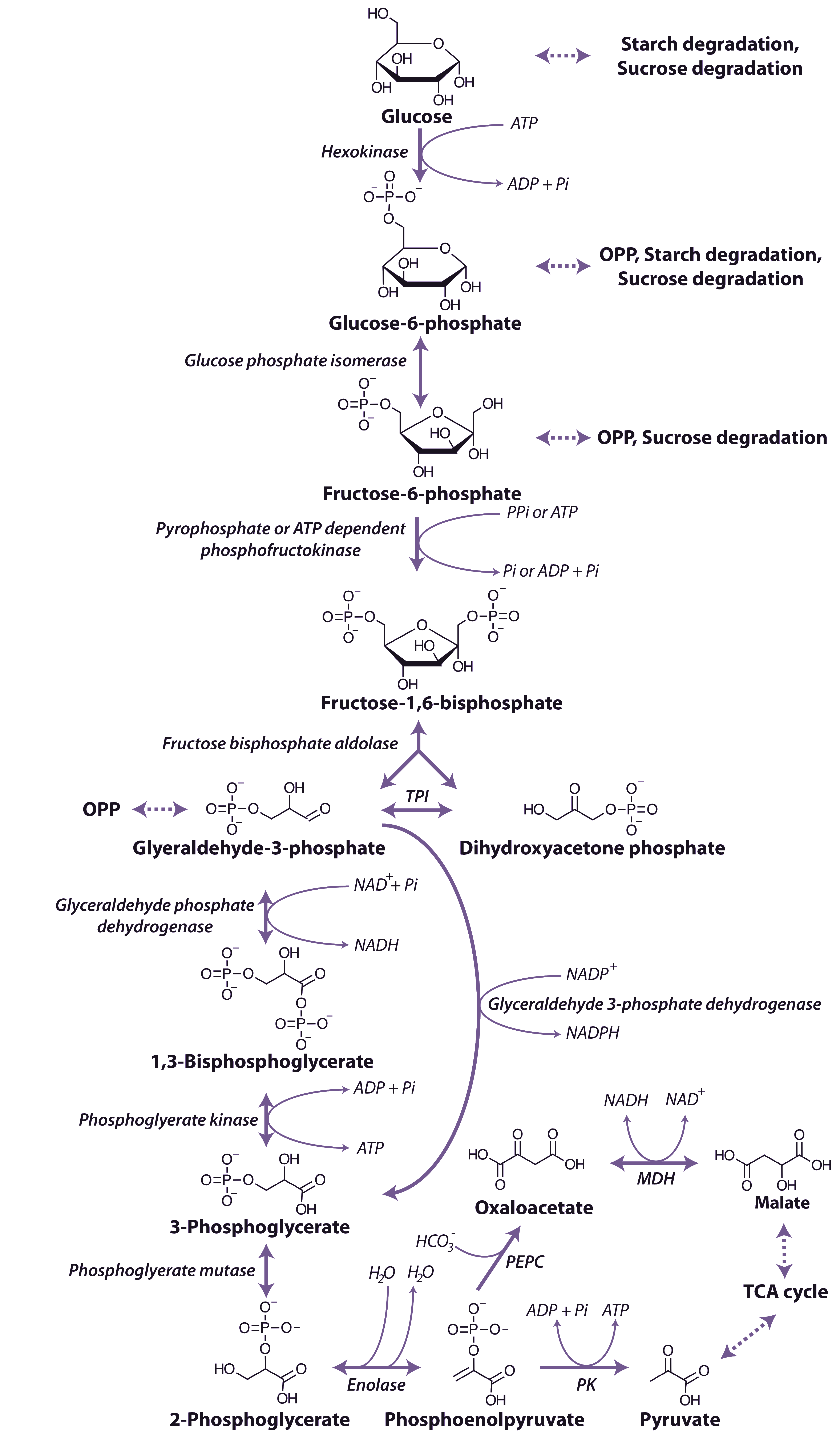
The glycolytic pathway involves the oxidation of the hexoses and hexose phosphates molecules produced from the breakdown of starch or sucrose to generate ATP, reductants and pyruvate (Figure 2.21).
Regulation of PFK and PPi-PFK is achieved by a combination of mechanisms, including pH, the concentration of substrates and effector metabolites and changes in subunit association. Phosphoenolpyruvate (PEP) is a potent inhibitor of both of PFK and PPi-PFK, inhibiting at µM concentrations and Pi can activate the cytoplasmic PFK, whereas the plastidic form is slightly inhibited by Pi. A number of other effectors of PFK have been identified including ADP, 3-phosphoglycerate and phosphoglycolate as well as it ability to accept ribonucleoside triphosphates other than ATP as the phosphate donor. PPi-PFK, has a catalytic potential higher than that of PFK and is strongly activated by Fructose-2,6-bisphosphate, but has no effect on PFK.
Fructose-1,6-bisphosphate is cleaved by fructose bisphosphate aldolase to form glyceraldehyde-3-phosphate and dihydroxyacetone phosphate, and these triose phosphates can be interconverted in a reaction catalysed by triose phosphate isomerase. Glyceraldehyde-3-phosphate is oxidised to 1,3-bisphosphoglycerate by a nicotinamide adenine dinucleotide (NAD+)-dependent glyceraldehyde 3-P dehydrogenase. Glyceraldehyde 3-P dehydrogenase is sensitive to inhibition by the reduced pyridine nucleotide cofactor (NADH), which must be reoxidised to maintain the flux through the glycolytic pathway. A phosphate group is then transferred from 1,3-bisphosphoglycerate to ADP forming ATP and 3-phosphogylcerate by phosphoglycerate kinase. In the cytosol a bypass is present that can convert glyceraldehyde-3-phosphate directly to 3-phosphoglycerate without phosphorylation by a non-phosphorylating NADP dependent glyceraldehyde 3-phosphate dehydrogenase. The resulting 3-phosphoglycerate is then converted to phosphenolpyruvate (PEP) by the action of phosphoglycerate mutase and then enolase.
The end-products of glycolytic reactions in the cytosol are determined by the relative activities of the two enzymes that can utilise PEP as a substrate: pyruvate kinase, which forms pyruvate and a molecule of ATP, and PEP carboxylase, which forms oxaloacetate (Figure 2.21). Both of these reactions are essentially irreversible and there are fine controls that regulate the partitioning of PEP between these reactions. Pyruvate kinase is controlled post translationally by a partial C-terminal truncation which may yield altered regulatory properties and a phosphorylation and ubiquitin conjugation that targets the protein to the 26S proteasome for complete degradation, it is also inhibited by ATP. Whereas PEP carboxylase is inhibited by malate and thus its regulation is independent of cell energy status. The sensitivity of PEP carboxylase to malate is regulated by phosphorylation of a N-terminal serine of the enzyme, with the phosphorylated form less sensitive to malate inhibition. Oxaloacetate is then reduced by malate dehydrogenase to malate which, along with pyruvate, can be taken up into mitochondria and metabolised further in the TCA cycle (see below). The reduction of oxaloacetate in the cytosol could provide a cytosolic mechanism for oxidising NADH formed by glyceraldehyde 3-P dehydrogenase (Figure 2.21).
Another level of regulation of components of glycolysis is their physical location within the plant cell. Under conditions of high respiratory activity, a greater proportion of the cytosolic enzymes of glycolysis are present on the surface of mitochondria. In contrast when respiration is experimentally inhibited, a decrease in the association of glycolytic enzymes with the mitochondria is observed. It is likely that the glycolytic enzymes associate dynamically with mitochondria to support respiration and that this association restricts the use of glycolytic intermediates by competing metabolic pathways.
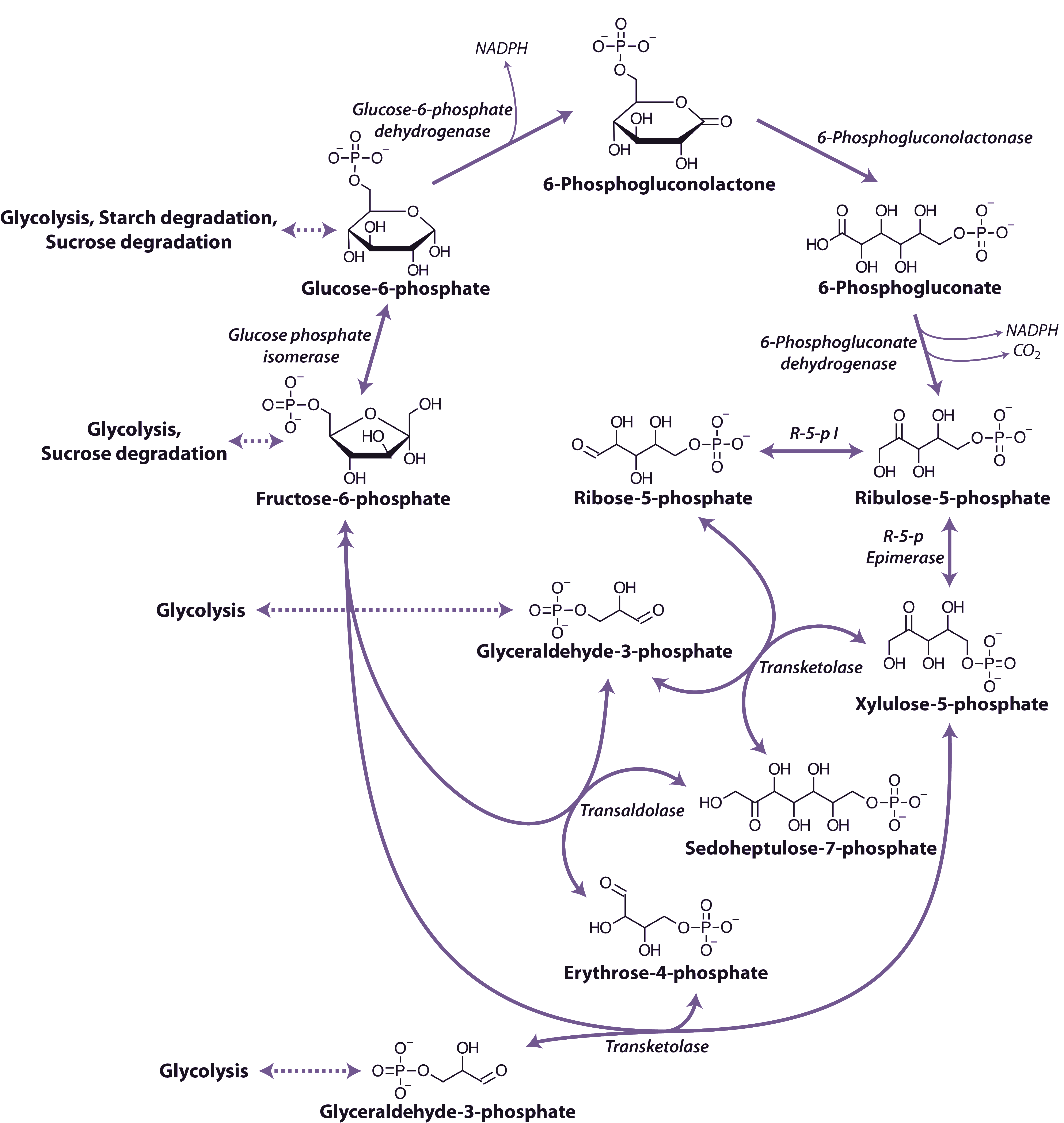
An alternative route for the breakdown of glucose-6-phosphate is provided by the oxidative pentose phosphate pathway (OPP) (Figure 2.22). This pathway functions mainly to generate reductant (i.e. NADPH) for biosynthetic processes including the assimilation of inorganic nitrogen and fatty acid biosynthesis and to maintain redox potential to protect against oxidative stress. In addition, the reversible oxidative section of the pathway is the source of carbon skeletons for the synthesis of a number of compounds. For example ribose-5-phosphate provides the ribosyl moiety of nucleotides and is a precursor for the biosynthesis of purine skeletons and erythrose-4-phosphate, which is the precursor for the biosynthesis of aromatic amino acids by the shikimic acid pathway.
The pathway begins with the dehydrogenation of glucose-6-phosphate catalyzed by glucose-6-phosphate dehydrogenase to produce 6-phosphoglucolactone and is the first step of the oxidative phase of the pathway. The 6-phosphoglucolactone is then hydrolysed to 6-phosphogluconate by 6-phosphogluconolactonase and then undergoes oxidative decarboxylation by 6-phosphogluconate dehydrogenase to produce ribulose-5-phosphate in the final step of the oxidative phase. Overall this phase of the pathway produces two molecules of NADPH from the conversion of glucose-6-phosphate to ribulose-5-phosphate. The non-oxidative phase begins with the reaction of ribulose-5-phosphate with either ribulose-5-phosphate isomerase or ribulose-5-phosphate epimerase followed by a series of reactions catalyzed by transaldolase and transketolase. These reactions result in the production of two molecules of fructose-6-phosphate and one glyceraldehyde 3-phosphate. The glyceraldehyde 3-phosphate and fructose-6-phosphate in the oxidative pentose phosphate pathway may be exchanged with enzymes of glycolysis.
As with glycolysis, reactions of the pentose phosphate pathway are catalysed by different isoforms of the enzymes that occur either in the cytosol or in plastids. Although transketolase and transaldolase may be absent from the cytosol of some species, the activity is maintained by phosphate translocator proteins on the plastid inner-envelope membrane that have the capacity to translocate sugar phosphates.
Organic acids such as pyruvate and malate produced in the cytosol by processes described above are further oxidised in mitochondria by the tricarboxylic acid (TCA) cycle and subsequent respiratory chain. Energy released by this oxidation is used to synthesise ATP which is then exported to the cytosol for use in biosynthesis and growth.
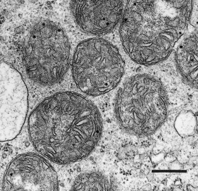
Plant mitochondria (Figure 2.23) are typically double-membrane organelles where the inner membrane is invaginated to form folds known as cristae to increase the surface area of the membrane. The outer membrane contains relatively few proteins (<100) and is permeable to most small compounds (< Mr=5 kDa) due to the presence of the pore-forming protein VDAC (voltage dependent anion channel) which is a member of the porin family of ion channels. The inner membrane is the main permeability barrier of the organelle and controls the movement of molecules by means of a series of carrier proteins many of which are members of mitochondrial substrate carrier family (MSCF). The inner membrane also houses the large complexes that carry out oxidative phosphorylation and encloses the soluble matrix which contains the enzymes of the TCA cycle and many other soluble proteins involved in a myriad of mitochondrial functions.
Mitochondria are semi-autonomous organelles with their own DNA and protein synthesis machinery. However, the mitochondrial genome encodes only a small portion of the proteins which make up the mitochondrion; the rest are encoded on nuclear genes and synthesised in the cytosol. These proteins are the transported into the mitochondrion by the protein import machinery and assembled with the mitochondrially synthesised subunits to form the large respiratory complexes. The number of mitochondria per cell varies with tissue type (from a few hundred in mature differentiated tissue to some thousands in specialised cells). Understandably, more active cells, with high energy demands, such as those in growing meristems are generally equipped with larger numbers of mitochondria per unit cell volume, and consequently show faster respiration rates.
Two substrates are produced from glycolytic PEP for oxidation in mitochondria: malate and pyruvate (Figure 2.21). These compounds are thought to be the most abundant mitochondrial substrates in vivo. However, amino acids may also serve as substrates for mitochondrial respiration in some tissues, particularly in seeds rich in stored protein or under conditions of sugar depletion such as extended darkness, shading and senescence. β-oxidation of fatty acids typically does not occur in plant mitochondria, this oxidation is principally carried out in peroxisomes in plants.
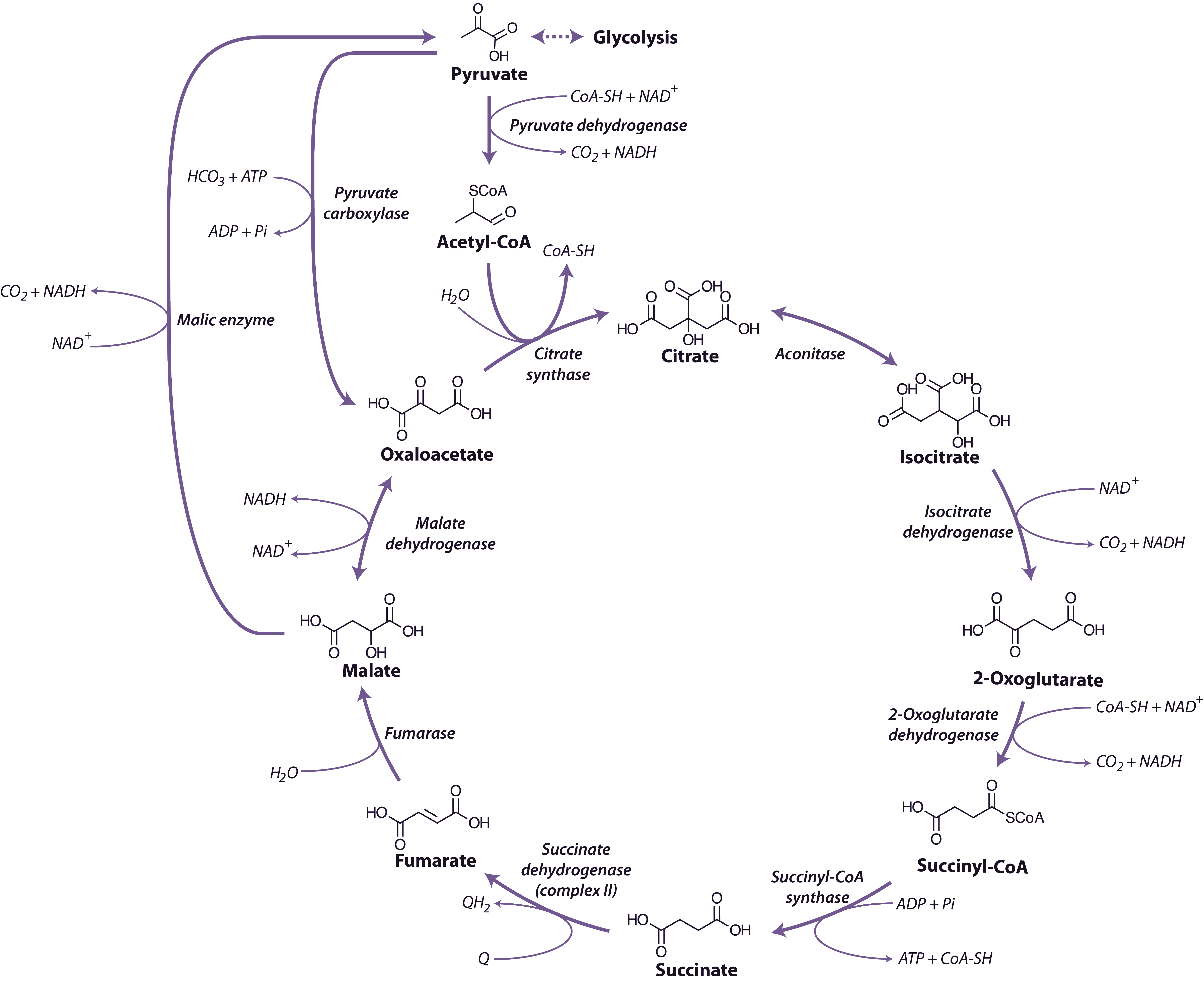
Malate and pyruvate enter the mitochondrial matrix across the inner membrane via separate carriers. Malate is then oxidised by either malate dehydrogenase (a separate enzyme isoform from that in the cytosol), which yields oxaloacetate (OAA) and reduced nicotinamide adenine dinucleotide (NADH), or NAD+-linked malic enzyme, which yields pyruvate and NADH and releases CO2 (Figure 2.24). Cytosolic pyruvate carboxylase is an alternative means of providing substrate to mitochondria by combining pyruvate with HCO3 to yield OAA that can then be imported into mitochondria.
Pyruvate formed either from malate and malic enzyme or transported directly from the cytosol is oxidised inside mitochondria by the pyruvate dehydrogenase complex (PDC) to form CO2, acetyl-CoA and NADH. This enzyme, which requires coenzyme A, thiamine pyrophosphate and lipoic acid as cofactors, effectively links the TCA cycle to glycolysis. PDC comprises three enzymes E1 (2-oxo acid dehydrogenase), E2 (acyltransferase) and E3 (lipoamide dehydrogenase). This complex is regulated by phosphorylation of the E1 subunit, lowering PDC activity in the day and increasing PDC activity at night. Pyruvate dehydrogenase is also subject to feedback inhibition from acetyl-CoA and NADH.
The TCA cycle begins with the condensation of acetyl-CoA and OAA, to form the six-carbon molecule citrate and the release coenzyme A (CoA) (Figure 2.24) in a reaction catalysed by citrate synthase. Aconitase catalyses the next step, converting citrate to isocitrate in a two-step reaction (dehydration/hydration) with cis-aconitate as an intermediate.
NAD-linked isocitrate dehydrogenase then oxidatively decarboxylates isocitrate to form CO2 and 2-oxoglutarate, and reduce NAD+ to NADH. The 2-oxoglutarate formed is also oxidatively decarboxylated to succinyl-CoA in a reaction catalysed by the enzyme 2-oxoglutarate dehydrogenase. This enzyme complex has similarities to pyruvate dehydrogenase and its reaction is analogous to the formation of acetyl-CoA from pyruvate by pyruvate dehydrogenase. The reaction mechanisms are also very similar, with 3 subunit enzymes, but 2-oxoglutarate dehydrogenase is not subject to the phosphorylation control that regulates pyruvate dehydrogenase. Succinyl-CoA synthase then catalyses the conversion of succinyl-CoA to succinate, with the concomitant phosphorylation of ADP to ATP, the only substrate-level phosphorylation step in the mitochondrion. This enzyme in plants differs from its mammalian counterpart in that it is specific for ADP rather than GDP.
Succinate dehydrogenase (SDH, Complex II), which catalyses the oxidation of succinate to fumarate, is the only membrane-bound enzyme of the TCA cycle and is part of the respiratory electron transfer chain (Figure 2.25). SDH is a large complex consisting of four core subunits, as well as number other associated subunits.
Fumarase catalyses the hydration of fumarate to malate followed by malate dehydrogenase that catalyses the final step of the TCA cycle, oxidising malate to OAA and producing NADH. The reaction is freely reversible, although the equilibrium constant strongly favours the reduction of OAA, necessitating rapid turnover of OAA and NADH to maintain this reaction in a forward direction.
Overall, during one turn of the cycle, three carbons of pyruvate are released as CO2, one molecule of ATP is formed directly, and four NADH and one FADH2 are produced. The strong reductants are oxidised in the respiratory chain to reduce O2 and produce ATP. Although most of the TCA cycle enzymes in plant mitochondria are NAD linked, NADP-dependent isoforms of isocitrate and malate dehydrogenases also exist, and these may play a role in a protective reductive cycle in the matrix.
Regulation of carbon flux through the TCA cycle probably occurs via phosphorylation/dephosphorylation of pyruvate dehydrogenase, which will depend in turn on mitochondrial energy status and feedback inhibition of various enzymes by NADH and acetyl-CoA. The rate of cycle turnover thus depends on the rate of electron flow through the respiratory chain (to reoxidise NADH) and the utilisation of ATP in the cell to provide ADP for substrate level and oxidative phoshorylation. TCA cycle turnover will also depend on the rate of substrate provision by reactions in chloroplasts and cytosol. A number of studies of TCA cycle mutants have demonstrated the wide impact these enzymes have not simply on TCA cycle function but as steps for the delivery of organic acids for other processes in plant cells such a photosynthetic performance, plant biomass, root growth, photorespiration, nitrogen assimilation, amino acid metabolism, and stomatal function.
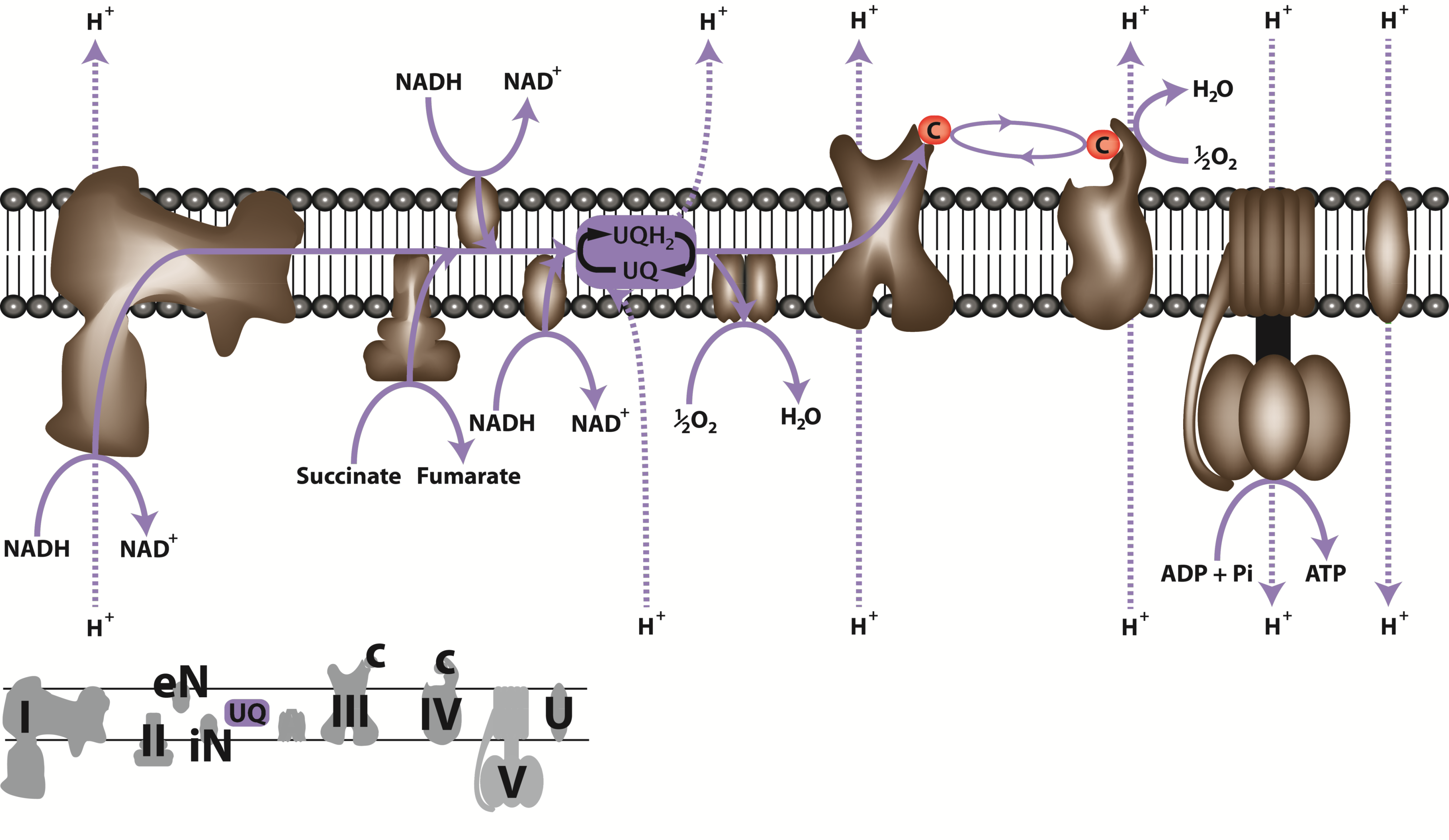
The respiratory electron transfer chain (ETC) of mitochondria consists of a series of large membrane-bound protein complexes (Complexes I, II, III, IV) which together with a small lipid ubiquinone (UQ) and the small protein cytochrome c catalyse the transfer of electrons from NADH and succinate to O2, forming H2O (Figure 2.25). Electron flow from NADH and succinate to oxygen is coupled to proton translocation out of the matrix to the intermembrane space which establishes a proton electrochemical gradient (DµH+) across the inner membrane that is used to drive phosphorylation of ADP to form ATP by the F1FO ATP synthase (Complex V, Figure 2.25).
NADH-UQ oxidoreductase, is responsible for the oxidation of matrix NADH and reduction of ubiquinone (UQ) in the inner mitochondrial membrane (Figure 2.25). In plants it is a large multi-subunit complex composed of 49 subunits, up to ten of which are synthesised in the mitochondrion whilst the others are imported from the cytosol. One of the subunits, a 50 kDa protein, contains flavin mononucleotide as a cofactor and is the dehydrogenase which oxidises NADH and passes electrons to iron-sulphur containing subunits of the complex, and eventually to ubiquinone. The passage of electrons through the complex is accompanied by H+ translocation across the membrane. Complex I is inhibited specifically by the flavonoid rotenone and its analogues. The NADH-binding site is exposed to the matrix and the complex oxidises NADH produced by the TCA cycle and other NAD-linked enzymes (Figure 2.25). Studies of mutations of CI subunits have shown that plants can survive without CI due to the activity of alternative NAD(P)H dehydrogenases (see below). Such mutants have a variety of interesting phenotypes including viral infection tolerance, prolonged hydration under water-deficient conditions and altered organic and amino acid concentrations. Using a series of Arabidopsis CI subunit knockout mutants, a number membrane arm subcomplexes (of 200, 400, 450 and 650 kDa) have been identified using BN-PAGE and antibodies. It is proposed that at least some of these subcomplexes may be assembly intermediates during CI formation, and these are seen to accumulate when specific subunits are absent.
Succinate dehydrogenase, is an enzyme of both the respiratory ETC and the TCA cycle (see above) (Figure 2.25). It is composed of four core subunits: a flavoprotein (SDHI), an iron-sulphur subunit (SDH2) and two membrane anchor subunits (SDH3 and SDH4) in most organisms. In plants, the purification of the complex has revealed the common core subunits, but also additional proteins of unknown function that co-migrate with the complex. All SDH subunits are encoded in the nuclear genome in Arabidopsis. SDH contains FeS and flavin centres which participate in electron transfer from succinate to ubiquinone. Unlike complex I, complex II does not pump H+ and succinate oxidation is therefore linked to the synthesis of less ATP per O2 reduced (see below). Malonate, an analogue of succinate, is a strong competitive inhibitor of succinate dehydrogenase. Knockout mutants of the SDH1 gene have been shown to be embryo lethal, but knockdown of SDH1 and SDH2 leads a array of phenotypes including altered stomatal aperture, mitochondrial ROS production and nitrogen use efficiency. SDHAF1 and SDHAF2 assist in CII assembly in plants and knockdown of the SDHAF2 homolog lowers SDH assembly and reduces root growth.
Ubiquinone-cytochrome c oxidoreductase or the cytochrome b/c1 complex as it is sometimes known, contains 10 subunits including a number of bifunctional core proteins. These proteins act both in CIII function and as a matrix processing peptidase, removing targeting presequences from imported matrix synthesized proteins (Figure 2.25). A single subunit of this complex, cytochrome b, is encoded by the plant mitochondrial genome, whilst the others are encoded by the nuclear genome. It contains two b-type cytochromes, b566 and b562, cytochrome c1, an iron-sulpher protein named the Rieske iron–sulphur protein and several other polypeptides. Electron flow from ubiquinol to cytochrome c is accompanied by the translocation H+ across the membrane, via the so-called Q cycle. Various inhibitors of complex III have been discovered, with antimycin A and myxothiazol most widely used in research. The assembly of CIII is modular and includes an early core subcomplex, a late core subcomplex and the final dimeric CIII. Approximately 13 assembly factors implicated in aiding one or more of the different stages of CIII assembly in yeast, however little is known about CIII assembly or functional assembly factors in plants.
Cytochrome c oxidase is the final step of electron transfer of the classical ETC. As the name implies, cytochrome c oxidase accepts electrons from cytochrome c and transfers them to O2 which is reduced to form H2O. Purification of CIV in plants has identified a complex containing 14 protein subunits (Figure 2.25). Eight of these proteins are homologous to known CIV subunits from other organisms, together with a further six proteins that are probably plant specific. Two cytochrome haem centres, a and a3, and two copper atoms make up its redox active components and like complex I, cytochrome oxidase is a proton pump. Cytochrome c oxidase is sensitive to a number of inhibitors, the best known of which are carbon monoxide and cyanide. Plants, however, show resistance to both carbon monoxide and cyanide because they are equipped with an alternative oxidase (see below). Studies of human and yeast CIV has shown an assembly pathway that involves the sequential incorporation of CIV subunits, initiated by subunit 1 and assisted by over 40 assembly factors. Research investigating the plant homolog of the yeast assembly factor COX19 has found it is capable of complementing the yeast cox19 null mutant. This suggests it might play a role in the biogenesis of plant cytochrome c oxidase or in the replacement of damaged forms of the enzyme. However, our knowledge of the assembly of CIV in plants is still incomplete.
These large multi-subunit complexes (I, II, III, IV) of the respiratory ETC chain are embedded in the inner mitochondrial membrane by virtue of their hydrophobic subunits, and interact with one another via two smaller molecules: ubiquinone and cytochrome c. The lipid-soluble ubiquinone also known as coenzyme Q10 is a small mobile electron carrier which moves rapidly along and across the membrane, and participates in H+ transport across the membrane via the Q-cycle as well as shuttling electrons from complexes I and II to complex III. Cytochrome c is a small haem-containing protein located on the outer surface of the inner membrane, which shuttles electrons between complexes III and IV. In this respect, the respiratory chain is similar in layout to the photosynthetic electron transport chain: three large complexes which communicate by a quinone and a small mobile protein (Cyt c or plastocyanin). However, orientation of components in the membrane is inverted and the net reaction catalysed is opposite to that in chloroplasts (Figure 1.12).
When electrons are transferred from NADH to O2, a large release of redox energy enables ATP formation in complex V of the respiratory chain (Figure 2.24). Energy release associated with electron transport is conserved by H+ translocation across the membrane to form a proton electrochemical gradient (ΔµH+) that has both an electrical membrane potential (Δψ) and a pH component (ΔµH+ = Δψ + ΔpH). This is known as the chemiosmotic theory and was originally proposed by Peter Mitchell in 1960s. In plant mitochondria, ΔµH+ exists mainly as a Δψ of ~150–200 mV, with a pH gradient (ΔpH) of ~0.2–0.5 units. ATP synthesis occurs as H+ move from a compartment of high potential (the intermembrane space) to one of low potential (the mitochondrial matrix) through the ATP synthase complex. Oxidation of NADH via the cytochrome pathway has three associated H+ translocation sites and is linked to synthesis of up to three ATP molecules for each molecule of NADH oxidized. By contrast, both succinate and alternative NADH oxidation by the rotenone-insensitive NADH dehydrogenases (see below) are linked to the synthesis of only two ATP molecules per NADH or succinate, as these events are associated with only two H+ pumping sites.
ATP synthase is a membrane-bound F1F0 type H+-ATP synthase that harnesses the ΔµH+ generated by the ETC to produce ATP. It is composed of a hydrophobic F0 component which channels protons through the inner mitochondrial membrane and also anchors the complex to the membrane and a hydrophilic F1 component which catalyses ATP formation and protrudes into the matrix. The core subunits of the enzyme are highly conserved in both prokaryotic and eukaryotic organisms. In plants, the majority of mitochondrial F1 subunits are encoded in the nucleus and translated in the cytosol before being imported into the mitochondria (including α, β, γ and ε subunits), while most of the F0 subunits are encoded in the plant mitochondrial genome and translated in the mitochondrial matrix (including a, b, c and A6L subunits). The reaction mechanism of the ATP synthase is known as the three-site alternating binding site mechanism. According to this model, F1 has three nucleotide-binding sites which can exist in three configurations: one with loosely bound nucleotides, one with tightly bound nucleotides and the third in a nucleotide-free state. H+ movement through F0 results in rotation of F1, causing a conformational change during which the site with loosely bound ADP and Pi is converted to one which binds them tightly in a hydrophobic pocket in which ATP synthesis occurs. Further H+ movement then causes another rotation of F1 and the ATP binding site is exposed and releases the nucleotide. In the meantime, the other nucleotide-binding sites are undergoing similar changes, with ADP and Pi being bound and converted to ATP. Thus H+ translocation drives the three sites through three different configurations and the main expenditure of energy is in the induction of a conformational change that releases tightly bound ATP, rather than in ATP synthesis itself. The F0 complex also contains a protein known as the oligomycin-sensitivity-conferring protein (OSCP) because it binds the antibiotic oligomycin that prevents H+ translocation through F0 and inhibits ATP synthesis. Therefore, adding oligomycin to mitochondria oxidising a substrate in the presence of ADP restricts O2 uptake. Generally knockouts of ATP synthase core subunits are lethal in plants, however inducible knockdowns have enabled investigations into the tissue-specific phenotypes incurred by slowing the rates of mitochondrial ADP:ATP cycling at a number of different developmental stages. It has been proposed that the assembly of plant CV comprises of three steps, the first being the formation of a rapidly turned over F1 subcomplex in the matrix, followed by an intermediate stage where F1 associates with the inner membrane and still turns over at a fast rate, and then finally a union of F1 with FO to form functional CV. A number of assembly factors (Atp10, Atp11, Atp12, Atp22, Atp23 and Fmc1) have been discovered for yeast ATP synthase, however, a detailed study of the presence and conservation of CV assembly factors in plants has not been undertaken.
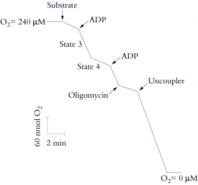
Electron transport through the respiratory chain, and therefore rate of O2 uptake, is controlled by availability of ADP and Pi, a phenomenon described as ‘respiratory control’. In the absence of ADP or Pi, the proton pore of ATP synthase is blocked and a ΔµH+ builds up to a point where it restricts further H+ translocation across the inner membrane. Since electron transport is functionally linked to H+ translocation, this elevated ΔµH+ will also restrict O2 consumption. That outcome is easily seen with isolated mitochondria (Figure 2.26) where O2 uptake is stimulated by adding ADP (‘State 3’ respiration). When all of the added ADP has been consumed, O2 uptake decreases again (‘State 4’). In steady state, the rate of electron flow is determined by the rate of flow of H+ back across the membrane: when ADP and Pi are available the backflow is rapid and occurs via ATP synthase; in the absence of these compounds, backflow is by slow diffusion through the membrane.
The ratio of State 3 to State 4 (the respiratory control ratio) is thus an indication of coupling between ADP phosphorylation and electron transport. Larger values represent tighter coupling. The proton leak can be dramatically stimulated by some compounds which act as protonophores or proton channels; these compounds collapse the ΔµH+ and increase O2 uptake up to the State 3 rate (Figure 2.26). However, no ATP is formed and these compounds are called uncouplers because they uncouple the linked processes of electron transport and phosphorylation.
Plant mitochondria have a respiratory chain which is more complicated than that of animals and contains alternative NADH dehydrogenases, alternative oxidases which catalyse cyanide-insensitive O2 consumption and uncoupling proteins that acts to dissipate the ΔµH+. The alternative NADH dehydrogenases and alternative oxidase do not translocate protons and therefore are not linked to ATP synthesis; they are often referred to as the non-phosphorylating bypasses of the plant respiratory chain. These pathways were initially identified in plant mitochondria as they are able to continue to respire in the presence of the CIV inhibitor, cyanide and the CI inhibitor, rotenone and by their ability to exhibit natively uncoupled respiration in the absence of an ADP source.
Cyanide-insensitive respiration is catalysed by the alternative oxidase (AOX). This alternative terminal oxidase is a diiron quinol oxidase that branches from the classical respiratory chain at UQ and reduces oxygen to water without an associated proton translocation. The oxidase exists in mitochondria as a dimer which can be inactivated by covalent linkage via disulphide bonds. The reduced enzyme is stimulated allosterically by pyruvate and some other 2-oxo acids (such as glyoxylate), which interact directly with the oxidase. The exact role of AOX continues to be debated but it appears to play an antioxidant role in plant mitochondria. Research has shown it is actively induced by oxidative stress and the different genes for the oxidase have been shown to be both development- and tissue-specific. Knockout of AOX leads to reactive oxygen species and anthocyanin accumulation in the leaves exposed to a combination of high light and drought stress. AOX can be inhibited by hydroxamic acids such as n-propylgallate (nPG) and salicyl hydroxamic acid (SHAM).
Alternative NAD(P)H dehydrogenases have been shown to be present on both sides of the inner mitochondrial membrane. These type II NAD(P)H dehydrogenases oxidise external or cytosolic and matrix NADH and NADPH and are insensitive to the classical CI inhibitor rotenone. As with AOX, these enzymes do not translocate protons and therefore are not linked to ATP synthesis. The Arabidopsis genome contains seven genes encoding NAD(P)H dehydrogenases, although it appears that some of these isoforms are present in multiple subcellular compartments in addition to mitochondria.
Uncoupling proteins (UCPs) are members of the mitochondrial carrier family of proteins. They act to dissipate the ΔµH+ built up the ETC by transporting H+ back across the inner membrane uncoupling proton and electron transport. The reactive oxygen species superoxide activates UCPs and this suggests a possible mechanism for the engagement of this enzyme in vivo. Analysis of knockouts of UCP (AtUCP1) showed that its absence led to localized oxidative stress but did not impair the ability of the plant to withstand a wide range of abiotic stresses. However, knockout of UCP1 did limit the photorespiration rate of plants and led to a reduction in photosynthetic carbon assimilation. This suggests that the main role of UCP1 in leaves is to maintain the redox poise of the mitochondrial ETC to facilitate photosynthesis.
Respiration represents a substantial loss of carbon from a plant, and under adverse conditions can be as high as two-thirds of the carbon fixed daily in photosynthesis. Both the rate and the efficiency of respiration will therefore affect plant growth significantly. The overall process of respiration results in the release of a substantial amount of energy which may be harnessed for metabolic work. In theory, the energy released from the complete oxidation of one molecule of glucose to CO2 and H2O in respiratory reactions leads to the synthesis of 36 molecules of ATP. However, in plants, because there are alternative routes for respiration, this yield can be greatly reduced. Mechanisms for regulating respiration rates in whole plants remain unclear. Convention has it that the rate of respiration is matched to the energy demands of the cell through feed-back regulation of glycolysis and electron transport by cytosolic ATP/ADP. However, since plants have non-phosphorylating bypasses in their respiratory chain that are insensitive to ATP levels, and since PEP carboxylase and PFP might be involved in sucrose degradation, the situation in vivo is not so simple. For example, the rotenone-insensitive alternative NADH dehydrogenases requires high concentrations of NADH in the matrix before it can operate and seems to be active only when substrate is plentiful and electron flow through complex I is restricted by lack of ADP. Alternative oxidase activity also depends on carbon and ADP availability and its flux is very dependent on the degree of environmental stress of the plant. In other words, non-phosphorylating pathways act as carbon or reductant ‘overflows’ of the main respiratory pathway and will only be active in vivo when sugar levels are high and the glycolytic flux rapid, when the cytochrome chain is inhibited, or when the bypasses have been induced significantly during stress. In glycolysis, the interaction between environmental signals and key regulatory enzymes, as well as the role of PFP and its activator fructose-2,6-P2, will be important.
One way of viewing respiratory cost for plant growth and survival is by subdividing measured respiration into two components associated with (1) growth and (2) maintenance. This distinction is somewhat arbitrary, and these categories of process physiology must not be regarded as discrete sets of biochemical events. Such energy-dependent processes are all interconnected because ATP represents a universal energy currency for both, while a common pool of substrates is drawn upon in sustaining production of that ATP. Nevertheless, cells do vary in their respiratory efficiency, while genotype × environment interactions are also evident in both generation and utilisation of products from oxidative metabolism. The benefit of a high respiration rate is that more ATP is produced, which provides vital energy for growth of new tissue and defence processes, such as antioxidant activation, metabolite transport or production of resistant protein isoforms. However, the cost of high respiration rates is that carbon is expended on respiration instead of being allocated to synthesis of new tissue, therefore limiting growth capacity. Variation in respiration rate has implications for growth and resource use efficiency in plants during drought, temperature and salinity responses of plants.
Araujo WL, Nunes-Nesi A, Nikoloski Z, Sweetlove LJ, Fernie AR (2012) Metabolic control and regulation of the tricarboxylic acid cycle in photosynthetic and heterotrophic plant tissues. Plant Cell Environ 35: 1-21
Atkin OK, Macherel D (2009) The crucial role of plant mitochondria in orchestrating drought tolerance. Ann Bot 103: 581-597
Atkin OK, Tjoelker MG (2003) Thermal acclimation and the dynamic response of plant respiration to temperature. Trends Plant Sci 8: 343-351
Jacoby RP, Li L, Huang S, Pong Lee C, Millar AH, Taylor NL (2012) Mitochondrial composition, function and stress response in plants. Journal of Integrative Plant Biology 54: 887-906
Jacoby RP, Taylor NL, Millar AH (2011) The role of mitochondrial respiration in salinity tolerance. Trends Plant Sci 16: 614-623
Kruger NJ, von Schaewen A (2003) The oxidative pentose phosphate pathway: structure and organisation. Curr Opin Plant Biol 6: 236-246
Mannella CA (2008) Structural diversity of mitochondria functional implications. Mitochondria and Oxidative Stress in Neurodegenerative Disorders 1147: 171-179
Millar AH, Whelan J, Soole KL, Day DA (2011) Organization and regulation of mitochondrial respiration in plants. Annual Review of Plant Biology 62: 79-104
Moller IM (2001) Plant mitochondria and oxidative stress: electron transport, NADPH turnover, and metabolism of reactive oxygen species. Annu Rev Plant Physiol Plant Mol Biol 52: 561-591
Patrick JW, Botha FC, Birch RG (2013) Metabolic engineering of sugars and simple sugar derivatives in plants. Plant Biotechnol J 11: 142-156
Plaxton WC (1996) The organization and regulation of plant glycolysis. Annu Rev Plant Physiol Plant Mol Biol 47: 185-214
Stitt M (2013) Progress in understanding and engineering primary plant metabolism. Curr Opin Biotechnol 24: 229-238
Stitt M, Zeeman SC (2012) Starch turnover: pathways, regulation and role in growth. Curr Opin Plant Biol 15: 282-292
Streb S, Zeeman SC (2012) Starch metabolism in Arabidopsis. Arabidopsis Book 10: e0160
Sweetlove LJ, Beard KF, Nunes-Nesi A, Fernie AR, Ratcliffe RG (2010) Not just a circle: flux modes in the plant TCA cycle. Trends Plant Sci 15: 462-470
Sweetlove LJ, Fernie AR (2013) The spatial organization of metabolism within the plant cell. Annu Rev Plant Biol 64: 723-746
Taylor NL, Day DA, Millar AH (2004) Targets of stress-induced oxidative damage in plant mitochondria and their impact on cell carbon/nitrogen metabolism. Journal of Experimental Botany 55: 1-10
Tcherkez G, Mahe A, Hodges M (2011) (12)C/(13)C fractionations in plant primary metabolism. Trends Plant Sci 16: 499-506
Zeeman SC, Kossmann J, Smith AM (2010) Starch: its metabolism, evolution, and biotechnological modification in plants. Annu Rev Plant Biol 61: 209-234