3.5 - Water and nutrient transport through roots

3.0-Ch-Fig-3.46.jpeg

Figure 3.46. Transverse section of a mature maize root, showing many layers of cortical cells outside a distinctive suberised endodermis bounding the stele. Late metaxylem vessels with large diameters are the dominant feature of the stele. Note a hypodermal layer underlying the epidermis. Pink Toluidine blue staining characterises suberised cells. (Photograph courtesy A.W.R. Robards)

Water and nutrients travel radially across roots from the soil to the xylem vessels and from there, they travel axially to the shoot. Anatomical and morphological features determine how effective roots are at absorbing and transporting water and nutrients from the soil to the shoot. Root tissue needs to form a low resistance pathway for transport, but also minimise the loss of water to the soil under adverse conditions.

The complexity of the root tissue that water and nutrients must cross is shown below in Figure 3.46. Notable features are the many layers of cortical cells, and the stele containing large and small xylem vessels. The stele is enclosed by the endodermis, a cell layer that contains deposits of suberin and lignin in the anticlinal walls between cells, called a Casparian strip, which blocks diffusion of water and solutes from the cortex into the stele. All roots have an endodermis, except at the tip where they are still growing.

Figure 3.46 also shows a layer of cells beneath the epidermis, which is often suberised and called exodermis. The suberin forms in older roots of many species or under drought conditions. Water and solutes cannot flow across the suberised cells of the exodermis and are restricted to passage cells that are not suberised.

Across the cortex, water could flow along cells walls and through interstitial spaces, the so-called apoplastic route. Alternatively it could enter the epidermal or hypodermal layer and flow symplastically across the cortex. Another alternative is the transmembrane (or transcellular) route. These three pathways are described in the following section. The solutes in the soil solution may also take the same route, or move independently of water.

Radial root conductance to water varies with stage of root development. The radial pathway is considered as more limiting than the axial pathway, and in the younger part of the root where the endodermis and hypodermis are not suberised, the radial conductance to water is higher than when these cell layers are suberised.

Within the stele, water and solutes move symplastically from the endodermis to the adjacent layer of pericycle cells, which are not suberised, so water can then move apoplastically to the xylem vessels. Solutes would most likely stay within the symplasm and be transported via plasmodesmata to the xylem parenchyma cells, from where they are loaded into the xylem vessels. If the xylem vessels are mature and the plant is transpiring, the solutes are carried swiftly to the shoots. If the xylem vessels are immature, the solutes are later released after rupture of the end walls of the immature xylem elements.

3.5.1 - Radial pathways across roots

3.0-Ch-Fig-3.47.jpg

Figure 3.47. Sketch of a transverse section of a young root showing three proposed pathways of ion and water flow across roots. 1. Apoplastic, via cell walls. 2.  Transmembrane (transcellular), via cytoplasm within cells and crossing membranes via efflux and influx membrane transporters. 3. Symplastic, via cytoplasm all the way, crossing cell walls via plasmodesmata. (Drawing courtesy B. Berger, based on Plett and Moller 2010). Note that at this stage the endodermis is not suberised and has just a Casparian strip (red). Deposition of suberin in the endodermal wall, and the development of an exodermis, will restrict the apoplastic and transcellular pathways.

Water and ions may move through root tissues along one of three routes: symplastic, apoplastic, or transmembrane, the latter also known as the “transcellular pathway” (Figure 3.47). An update on the anatomical and molecular bases of these pathways in Arabidopsis is given by Barberon and Geldner (2014).

The proportion of water flowing through the different pathways undoubtedly varies with the plant’s rate of transpiration.

Apoplastic Flow

Water and soil solutes might move through the cell walls of the cortical cells until the endodermis, where they would cross the plasma membrane of the endodermal cells. Water would cross via aquaporins, and solutes via ion channels or transporters (see Section 3.6). This is possible in roots without a suberised hypodermis and when the endodermis is in the primary stage of development, as described in the following page. Entry of anions to deep layers of the cortex is likely to be restricted by charge repulsion from dissociated, negative carboxyl groups in cell walls (Donnan Free Space). In general, cations also pass through cell walls more readily than anions, particularly if many of the carboxyl groups in cell walls are not occupied by Ca2+ ions. Nonetheless, apoplastic flow of water through roots can sustain large ion fluxes during periods of high transpiration.

It is likely that the bulk of the soil water taken up by the plant moves in the apoplast across the cortex, but that solutes are taken up by epidermal or outer cortical cells and then move in the symplasm across the cortex.

When the endodermis is suberised, water and solutes cannot enter from the apoplast and instead are taken up into the adjacent cortical cells and move via plasmodesmata into the endodermis.

Symplastic Flow

Symplastic flow occurs when water or solutes are taken up by the epidermis or an outer cortical cell, and then subsequently cross the rest of the cortex via plasmodesmata. Plasmodesmata are extensions of the plasma membrane that cross cell walls and provide cytoplasmic continuity between cells, allowing neighbouring cells to communicate and exchange materials more freely. Water and ions can move through the cortex via a series of plasmodesmal connections, thereby remaining in the cytoplasm until reaching the stele. Rate of water and nutrient transport is largely regulated by plasmodesmal resistance, which depends on size and frequency of plasmodesmata in the cell membranes. Alternatively, water and some ions enter vacuoles and are therefore, subject to transport properties of the tonoplast.

Transmembrane (transcellular) Flow

A third possibility is the transmembrane or transcellular route, in which water and nutrients cross membranes, passing repeatedly between symplasm and apoplasm as they are transported from one cell to the next. For nutrients, this form of transport is facilitated by carriers that are distributed in a polar fashion, with influx carriers located on the outer walls and efflux carries on the inner walls. Water transport across cell membranes is via aquaporins for which there is no “rectification”, or intrinsic directionality of transport. They are therefore most likely to be distributed evenly along the plasma membrane.

This pathway may increase resistance to flow of water, but also provides a more effective mechanism for controlling the flow of solutes and water across the root than the purely symplastic route via plasmodesmata when only the first membrane crossing is available to control ion and water transport.

3.5.2 - Variable barriers: endodermis and exodermis

The epidermis and root hairs probably mediate most of the selective uptake of solutes from the soil solution. Nutrients are selectively taken up, and potential toxins excluded.  The cortical cell layers continue this selective uptake if solutes travel apoplastically towards the stele.

The endodermis is the innermost cortical layer that surrounds the central vasculature, and forms a barrier preventing water flow and free diffusion of solutes in the apoplast because of its Casparian strip and suberin lamellae. It thus, prevents soil solution moving into the stele without crossing a membrane, and also prevents water from within the stele being lost back to the soil at night.  Although, in some regions of the endodermis that are opposite xylem poles, water and solutes may bypass the endodermis through unsuberised “passage cells”. The endodermis also functions in structural support for the stele, particularly in drying soil, and minimises shrinkage or swelling of the cells of the stele. Its role in ion selectivity is minor compared to the cells of the cortex and epidermis.

In many plants, the cortical layer under the epidermis (the  hypodermis) develops Casparian bands and suberin lamellae, and develops into the exodermis. This forms another barrier for the apoplastic movement of water and solutes. Just like in the endodermis, some passage cells in the exodermis may be free of suberin and can take up water and solutes.

(a) Endodermis

An endodermis with a Casparian strip is a feature of roots of all land plants from ferns upwards. As the innermost cortical layer that surrounds the central vasculature of roots, the endodermis acts as a barrier to the free diffusion of solutes from the soil into the stele, and the stele into the soil. The protective functions range from efficient water and nutrient transport to defence against soil-borne pathogens. The genes and regulation mechanisms that drive the differentiation of this intricately structured barrier have been reviewed by Geldner (2013).

The development of the endodermis has three stages: the primary stage in which the Casparian strip forms, the second stage when suberin lamellae encases the entire endodermal cell, and the tertiary stage when the inner tangential walls thicken and a layer of cellulose is deposited over the suberin lamellae.

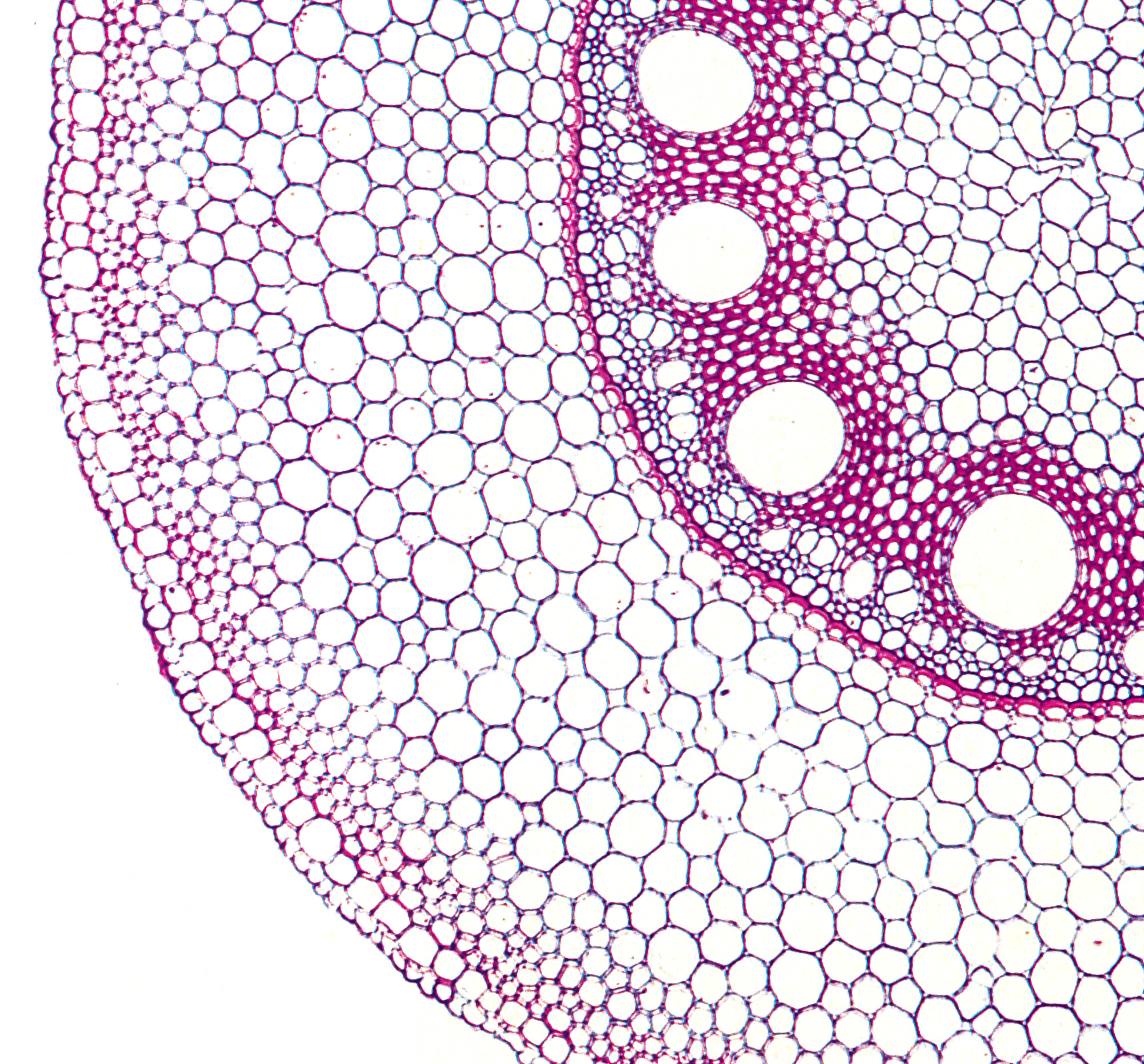
The Casparian strip is made of lignin and suberin and deposited as a ring in the radial walls of the endodermal cells, like a hoop around a barrel of beer. It reaches from the plasma membrane to the outermost part of the wall and adjoins adjacent cells so there are no air spaces between the cells at this point. It therefore blocks the flow of water through the cell walls. It differentiates as root cells mature about 5-10 mm from the tip, and so an entirely apoplastic pathway from soil to central stele can occur only in very young root parts or at sites where the Casparian strip is disrupted such as site of lateral root development, which starts at the pericycle, the cell layer beneath the endodermis.

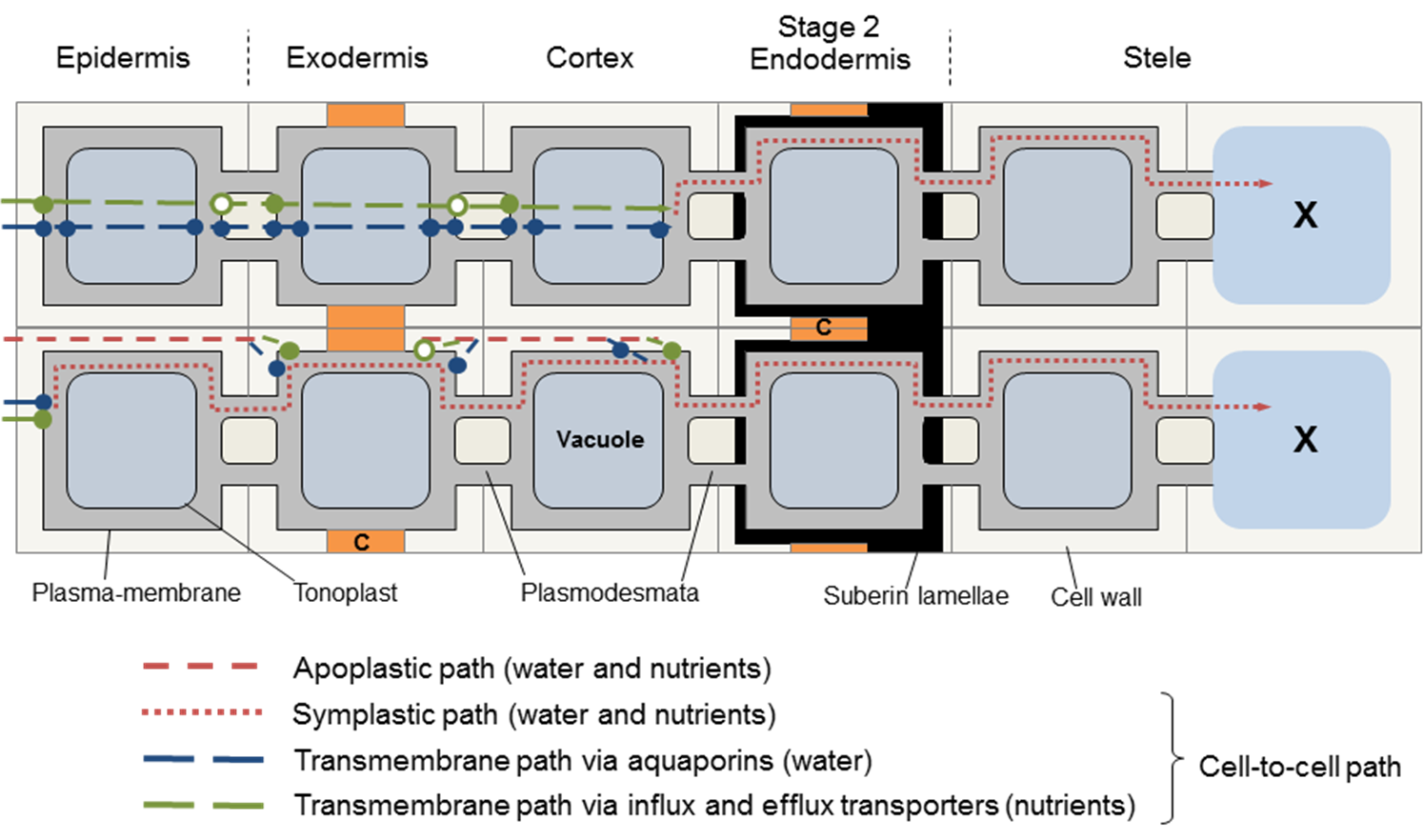
In the first stage of endodermis development, the Casparian strip in the primary wall prevents flow of water and solutes through the wall from inner cortex to stele. Any solutes remaining in the apoplastic water must enter the endodermis via membrane transporters. Water in the apoplast must enter the endodermis via aquaporins as illustrated in the lower pathway shown in Figure 3. 50. 

3.0-Ch-Fig-3.50.png

Figure 3.50. The alternative pathways for water and nutrient solute flow across roots with the endodermis at Stage 1 of development, with a primary wall with Casparian strip (c). The apoplastic pathway is blocked at this point, and water, along with any nutrients still in the walls, enters the endodermal cells via membrane transporters (lower panel). If the hypodermis also has a Casparian strip, water and nutrients must also enter via membrane transporters. Alternatively, water and nutrients can move via the symplastic (dotted red line, lower panel) or transmembrane pathways (upper panel). (Diagram courtesy H. Bramley)

In the second stage of development, the endodermis becomes suberised as the secondary walls develop, and suberin lamellae are formed all over the cell, underneath the primary wall that contains the Casparian strip. This occurs at varying distances from the root tip depending on species and is often induced under drought to form in younger parts of the root.

Suberin is a hydrophobic polymer, deposited in the secondary cell wall in lamellae. It therefore seals off the plasma membrane from solutes, as water and ion channels are sealed. So the transcellular and apoplastic pathways are curtailed, and water and solutes enter the endodermis only through plasmodesmata from neighbouring cells (Figure 3.51). Any function in selective control of particular nutrients into or out of the endodermal cell through the suberin lamellae is unclear.

3.0-Ch-Fig-3.51.png

Figure 3.51. Alternative pathways for water and nutrient flow across roots with a suberised (Stage 2) endodermis. A complete layer of suberin around the endodermis means that water and solutes can enter only via plasmodesmata from the inner cortical cells. (Diagram courtesy H. Bramley)

Some endodermal cells remain at the primary stage even late in root development, and are called passage cells.

Stage 3 of the endodermis involves further deposition of cellulosic wall material, further impeding flow of solution through walls.

(b) Exodermis

Exodermis is the name given to a hypodermis with Casparian strips and suberised lamellae.

Occurrence

Roots of most species form an exodermis with time. First, a Casparian band is laid down in the primary wall of the hypodermis, then all the wall is suberised especially the inner wall, as for the endodermis. Some cells in this hypodermal layer (‘passage cells’) remain unsuberised.

The development of the exodermis is very common in the plant kingdom (Perumalla et al. 1990). In a study of 180 angiosperm species, the great majority (89 %) showed a clearly suberised exodermis with Casparian strip. It was found in roots of primitive and advanced plant families, from hydrophytic, mesophytic and xerophytic habitats, but was lacking in some Poaceae. It was notably absent in oat, barley and wheat (Perumalla et al. 1990), but generalizations within cereals cannot be made as subsequently maize was found to have an exodermis (Hose et al. 2001),

The Casparian band can develop close to the root tip. For example, in aeroponically grown maize a complete exodermal layer formed 30 mm above the root tip. In roots elongating more slowly due to abiotic stress or low temperature, it can be found closer to the tip. The extent at which apoplastic barriers form depends on the stage of development of the root system and also the habitat: drought, waterlogging, salinity, nutrient deficiency or toxicity may strongly influence the degree of suberisation (Hose et al. 2001).

Permeability to water and solutes

3.0-Ch-Fig-3.54.png

Figure 3.54. Schematic diagram of a longitudinal section through a root indicating the stage at which the critical structures in radial and axial transport of water develop. Not to scale. (Diagram courtesy H. Bramley)

When an exodermis forms, it also imposes a restriction to radial transport. Complete layers of suberin constrain water and solute flow only via plasmodesmata from the epidermis and inner cortical cells.  Whether or not it can be considered as a barrier depends on the degree of suberisation and the number of passage cells within it. Its properties as a barrier are variable (Hose et al. 2001).

The exodermis represents a resistance to the radial flow of both water and solutes, much like the endodermis in Stage 1 development. It restricts radial apoplasmic movement and may also restrict transmembrane transport of nutrients. Exodermal layers become functionally mature 20–120 mm from the apex, where lateral roots are initiated, and therefore constitute a barrier to apoplastic ion flow only in root zones where an endodermis is already present. In a similar way to the endodermis, maturation of an exodermis involves further deposition of suberin and cellulosic wall material, further impeding flow of solution through walls.

The exodermis is not totally impermeable to water or nutrients and may have a differential selectivity that varies with the environment. How much the root is sealed off depends on the type of exodermis: (1) uniform exodermis where the cells are uniform in shape (suberin deposition is patchy and develops late) or (2) dimorphic exodermis, which consists of long and short passage cells (the former of which are suberised). Suberin lamellae enclosing the long cells disrupt the cytoplasmic continuity through plasmodesmata and the cell dies, but suberin lamellae in the uniform-type exodermis do not affect plasmodesmata. Individual passage cells allow passage of solutes and water via uptake carriers, not via the apoplast as flow there is blocked by the Casparian strip. They have an active role in ion uptake and often become the only plasmalemma facing the soil solution especially when the epidermis dies. Some families (e.g. irises) have large numbers of passage cells while others have very few.

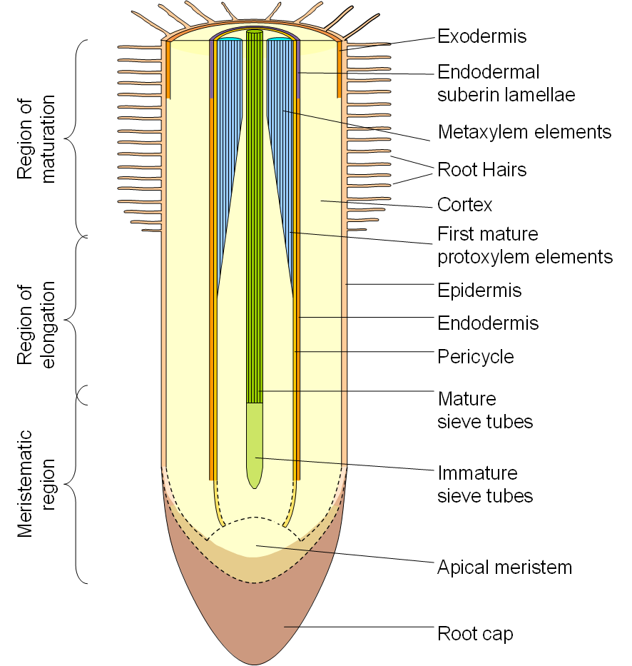
The diagram of a root to the right (Figure 3.54) indicates the stage of development at which the various structures are functional.

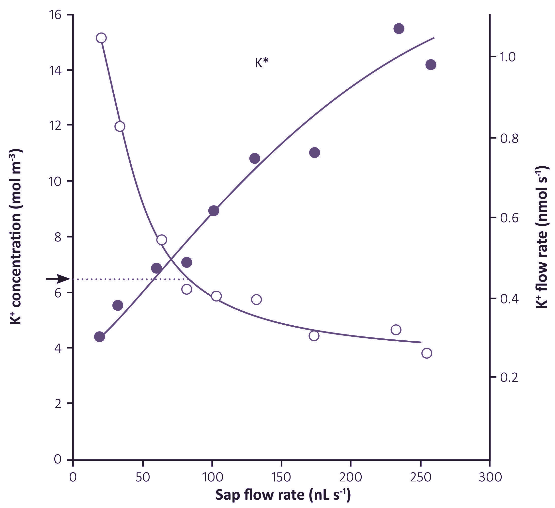
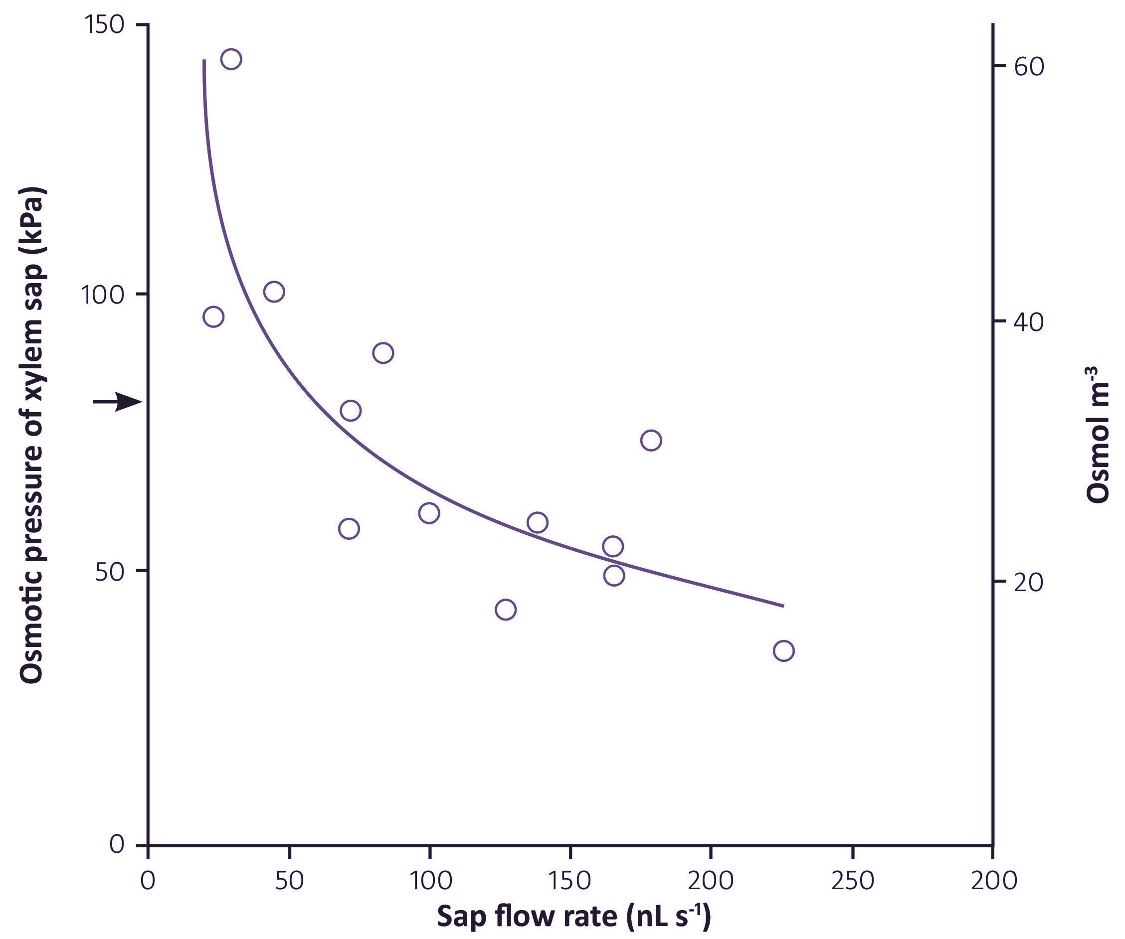
The anatomy of roots and the alternative pathways of water and solute flow across roots and into the xylem indicates that the relationship between water and nutrient flow to the shoots could be quite complex. Water may take different pathways than nutrients. The distribution of water in the different pathways depends not only on the age of the root, but may also vary with the flux of water moving through, that is, the rate of transpiration.

3.5.3 - Relation between water and nutrient flux

3.0-Ch-Fig-3.55.jpeg

Figure 3.55. Effect of volume flux (sap flow rate) on K+ concentration and K+ flux in xylem sap from barley. Arrow indicates the external K+ concentration, and dotted line indicates the range of sap flow rate for which  K+ in the sap was at a higher concentration than the external solution. (Munns, 1985)


The various pathways taken by water and nutrients, and the environmental factors that influence them, are not well understood. The fundamental processes governing the relationship between water and nutrient flow through roots are complex. One thing is clear: nutrients do not move passively with the transpiration stream. But neither is their movement entirely independent of it.

This section looks at the relation between water and solute fluxes to the shoot: how the rate of transpiration affects solute concentrations in the xylem, and how active solute transport affects water flow rates at night (root pressure).

(a) Effect of transpiration on solute flux in the xylem

Solute fluxes in the xylem usually respond to changes in transpiration rate, although the relationship is not proportional. The figure below (Figure 3.55) indicates an apparently strong relation between water flux and  K+ flux, but it must be noted that the flux of  K+ is already high at very low flow rates, increases linearly with moderate increases in flow rate, but then tapers off at high flow rates.

A similar relation has been found for other nutrients like nitrate.

The importance of transpiration in carrying nutrients to the shoot has long been debated. However, experimental evidence showed that transpiration was not necessary to get nutrients to the shoot, as growth rates and net nutrient transport rates were unaffected by humidity and other environmental conditions that would reduce transpiration (Tanner and Beevers 2001). Transpiration was not a prerequisite for long-distance transport of nutrients, as root pressure (see below) in the absence of transpiration can supply the shoot with the required nutrients.

(b) Effect of transpiration on solute concentration in the xylem

3.0-Ch-Fig-3.56.jpeg

Figure 3.56. Effect of transpiration rate on osmotic pressure (left hand axis) and the corresponding solute concentration (right hand axis) of xylem sap of young barley plants. Transpiration rates were imposed by varying vapour pressure deficit around the shoots and xylem sap was sampled by applying sufficient pressure to roots to cause a cut leaf tip to bleed. Arrow indicates the external osmotic pressure. (Munns and  Passioura, 1984)

Transpiration rate (the volume flux in the xylem) has a marked effect on the concentration of solutes in the xylem sap. Plants which are transpiring rapidly have a low concentration of nutrients in their xylem sap compared with slowly transpiring plants.  This relationship between concentration and volume flux holds for most nutrients, see example with K+ (Figure 3.55 above) in which increasing flow rates decreased the concentration of  K+ in xylem sap.

The data also shows that an apoplastic or diffusive uptake of K+ is not important because the concentration of K+ in the xylem was greater than the external K+ over a range of volume fluxes which produced an increasing flux (Figure 3.54). These data strongly suggest that ion concentrations in xylem sap are a result of either a single variable pump, or more likely of several sequential processes. The composition of the transpiration stream could be modified after the first passage across a membrane as it flows towards the stele or upwards through the stele by either active or passive ion fluxes from cortical or xylem parenchyma cells.  K+ could be pumped in (or diffuse in) at rates which increase with increasing volume flux.

Similar relationships occur between most solutes in the sap and transpiration rate, as shown in Figure 3.56.

This shows how much influence the transpiration rate has on the concentration of solutes in the xylem, and that concentrations in sap collected from exuding cut stumps are not typical of concentrations in transpiring plants.

(c) Root pressure

Plants that have had shoots removed have very concentrated xylem fluid, which exudes from the cut stumps under positive hydrostatic pressure from the roots (‘root pressure’). Root pressure is also responsible for the droplets of water seen on the margins of guttating leaves early in the morning (Figure 3.57).

3.0-Ch-Fig-3.57.jpeg

Figure 3.57. Guttation droplets from a eucalypt leaf, E. tetragona. (Photograph courtesy C. Hooper)

This occurs because nutrient uptake is an active process, independent of water uptake from the soil, and continues during the night – the rate of nutrient uptake varies little over 24 h. When nutrients are pumped into the stele, water flows in by osmosis, and the pressure builds up. Positive pressures of 30-300 kPa can be achieved in this way.

The Casparian strip and the suberisation of the endodermis is important as it provides a barrier to prevent back-flow of water and also structural support so that the root can contain a positive pressure at night. The pathways of nutrient and water flow across the root cortex may change at night, for instance the apoplastic pathway may be of lesser importance at low rates of transpiration. At zero transpiration induced by a root pressure probe and when the shoot is removed, pathways may be different again.

Root pressure is not just a phenomenon, it is an essential process responsible for moving nutrients to the shoot during the night when transpiration is low. It may also have a function in dissolving gas bubbles that might have caused cavitation in the xylem during the day.