2.2.8 - Crassulacean acid metabolism (CAM)

Joseph Holtum1, Klaus Winter2 and Barry Osmond3

1Centre for Tropical Biodiversity and Climate Change, James Cook University, Australia; 2Smithsonian Tropical Research Institute, Balboa, Ancón, Republic of Panama; 3School of Biological Sciences, University of Wollongong, and Research School of Biology, Australian National University, Australia

Crassulacean acid metabolism (CAM) is a water-conserving mode of photosynthesis that, like C4 photosynthesis, is a modification of the C3 photosynthetic pathway fitted with a CO2 concentrating mechanism (CCM) that can increase the [CO2] around ribulose bisphosphate carboxylase/oxygenase (Rubisco) by more than 10-fold and suppress photorespiration. The overall energy demand of the CAM pathway is only about 10% more than that of C3 photosynthesis, as costs of the CCM machinery are partially offset by reducing photorespiration.

In C4 plants, as explained earlier in Section 2.2.2, this CCM is most commonly achieved by an “in-line turbocharger” based on initial CO2 fixation by phosphoenolpyruvate carboxylase (PEPC) into C4 acids in the cytoplasm of outer mesophyll cells. These acids diffuse rapidly to adjacent relatively CO2-tight bundle-sheath cells (Figure 2.31 right) where CO2 is released again. High [CO2] builds up in this spatially separated compartment where it is refixed by Rubisco.

2.0-Ch-Fig-2.31.png

Figure 2.31 Leaf transverse sections of CAM versus C3 and C4 plants. Left: succulent CAM plant Kalanchoë daigremontiana. Centre: C3Atriplex hastata. Right: C4Atriplex spongiosa. Scanning electron  micrographs at similar magnification. (K. daigremontiana  image courtesy R.A. Balsamo and E.G. Uribe; Atriplex spp. images courtesy J. H. Troughton)

In CAM plants enzyme systems analogous to those in C4 plants achieve the same result through a “battery-like” dark accumulation of CO2 into the 2nd carboxyl group of malic acid (acidification phase) in the vacuole of large mesophyll cells (Figure 2.31 left). Malic acid can accumulate to very high concentrations, attaining concentrations of greater than 1 mole acid per litre in mesophyll cells of tropical tree-CAM plants (Clusia spp.). Indeed one can sometimes taste the acid, with acid taste-testing for the presence of CAM being possibly first recorded in Aloe sp. by Nehemiah Grew in 1682 and in field reports from India by Benjamin Heyne in 1815.

In the light, malic acid returns to the cytoplasm where it is rapidly decarboxylated (deacidification phase). The CO2 released, which accumulates to high internal [CO2] as stomata close, is refixed by Rubisco in chloroplasts of the same mesophyll cell where it is further assimilated by the photosynthetic carbon reduction (PCR) cycle (Figure 2.32).  

2.0-Ch-Fig-2.32.png

Figure 2.32  Schematic of the principal components of CAM, highlighting storage of malic acid in the vacuole (V) and the carbohydrate conundrum discussed later. (Diagram courtesy B. Osmond)

Ultimately three of the four carbons recovered from the malic acid must be stored as starch and/or sugars in order to provide to PEPC the C3 substrate required for CO2 uptake during the following night. The fourth carbon, effectively that obtained from the atmosphere, is available for growth. Deacidification may generate high [CO2] behind closed stomata but photorespiration is not completely abolished (Lüttge 2002) since photosynthesis also generates high internal [O2]. While exploring Lake Valencia in Venezuela in 1800, Alexander von Humboldt measured elevated [O2] in bubbles streaming from the cut base of presumably CAM Clusia leaves standing in water in the light.

Of course by closing stomata in the light CAM plants minimize water loss when evaporative demand is highest (von Caemmerer and Griffiths 2009). The biomass production per unit water utilized in CAM was 6 times higher than for C3 plants and 2 times higher than for C4 plants when plants exhibiting all three photosynthetic pathways were grown together in a garden outdoors (Winter et al. 2005). The attribute of water-use efficiency undoubtedly contributes significantly to the success of CAM photosynthesis in nature, with CAM species outnumbering C4 species by about two to one. The paradoxes of CAM, a mode of photosynthesis that involves stomatal opening and CO2 uptake during the dark, continue to inform many aspects of plant biochemistry, physiology, ecology and evolution. This article draws heavily on two recent reviews (Borland et al. 2011; Winter et al. 2015).

2.2.8.1 - Biochemical attributes distinctive to CAM

Although CAM and C4 photosynthesis share common enzyme machineries, the physiological bases of spatially-separated and time-separated CCMs are very different and involve complex suites of distinctive regulatory processes ranging from allosteric modulation of enzyme activities, through cell and organelle membrane metabolite transport systems, to long-term responses to stress. The resulting metabolism is rarely at steady state. It is thus helpful to reference the principal biochemical interacting components of CAM to the CO2 exchange patterns and the pool sizes of acidity and carbohydrates in the archetypical Kalanchoë daigremontiana as outlined in Figure 2.33.

2.0-Ch-Fig-2.33.png

Figure 2.33 Schematic outline of the phases of CAM, showing net CO2 exchange and malic acid and carbohydrate (glucan) metabolism in Kalanchoë daigremontiana leaves. (Diagram courtesy B. Osmond)

The four phases of CAM metabolism are:

  • Phase I - acidification in the dark (PEPC active and stomata open)
  • Phase II - a transitional phase with  stomata open and both carboxylases active
  • Phase III - deacidification (PEPC inhibited, Rubisco active and stomata closed)
  • Phase IV - C3 photosynthesis (stomata open, Rubisco active and PEPC inhibited)

Within these four phases, the distinctive underlying biochemistry of CAM involves the up-regulation of cytoplasmic PEPC activity during phase I in the dark. Up-regulation is catalysed by PEPC kinase which phosphorylates PEPC making it less sensitive to inhibition by malic acid as it accumulates in the vacuole. Towards night’s end, CO2 fixation by PEPC declines as its carbohydrate substrates are exhausted (Figure 2.33). PEPC kinase is degraded during phase II and PEPC becomes increasingly sensitive to malic acid (declining Ki malate; Figure 2.34). It remains inhibited throughout phases III and IV.

CAM also involves the up-regulation of Rubisco in the light by ATP-dependent Rubisco activase as photosynthetic electron transport (ETR) increases in phase II and is maintained throughout phases III and IV (Figure 2.34).

2.0-Ch-Fig-2.34.png

Figure 2.34 Regulation of enzyme activities in deacidification phases of CAM as photosynthetic ETR increases in the light. (Diagram based on K. Maxwell et al. Plant Physiol 121: 849-856, 1999)

Partitioning of carbohydrate metabolism occurs in the light to retain chloroplast starch or vacuolar sugars as substrates for the next nocturnal acidification phase (phase I) while diverting sugars for phloem transport and growth. In pineapple, for example, degradation of starch in the chloroplast may provide the substrate for PEPC despite the large diel turnover of soluble sugars. The complexity of this “conflict of interest” (Borland and Dodd 2002) in carbohydrate metabolism varies between CAM plants with different deacidification pathways.

Sophisticated interactions occur between metabolite transporters in membrane systems of the vacuole, mitochondria and chloroplasts. Many of these are unique to CAM but of 48 such transporters required to support known variations of CAM (including Clusia spp. that also accumulate citric acid) up until 2005, only 8 had been demonstrated in at least one species (Holtum et al. 2005).

When studied under constant conditions, many of the above distinctive biochemical processes in CAM exhibit circadian rhythms. The extent to which endogenous oscillators orchestrate the clearly interacting biochemical, physiological and environmental controls seems likely to remain a challenging area of research.

2.2.8.2 - Physiological attributes distinctive to CAM

2.0-Ch-Fig-2.35.png

Figure 2.35 Diel expansion growth of Opuntia oricola cladodes during drought in the desert biome of the Biosphere 2 Laboratory in Oracle Arizona USA measured by time-lapse photography. Heterogeneity of growth rate throughout the day is colour coded as red = 2.0% to blue = 0.5% per hour (Diagram by B. Osmond based on Gouws et al. Funct Plant Biol 32: 421-428, 2005)

Compared to the photosynthetic biochemistry and physiology in leaves of C3 and C4 plants, the 6% of taxa estimated to exhibit CAM (in at least 35 families and >400 genera) express it with staggering variety (Winter et al. 2015). That is, the distinctive biochemical attributes of CAM outlined above, derived from a handful of research-compliant leafy model species, are but the tip of an iceberg of what really qualifies as a CAM plant (Borland et al. 2011).

The following summary of some distinctive physiological attributes of CAM underscores this conundrum:

Biochemical and physiological determinants of stable isotopic composition of plants with CAM. Fixation of CO2 by PEPC and Rubisco in vitro show clearly different discriminations against the heavier, naturally occurring, non-radioactive (stable) 13C isotope of carbon when expressed as a \(\delta\)13C value. Thus total carbon in C4 plants reflects a small discrimination against 13C resulting in \(\delta\)13C values of about –12.5 ‰, with more negative values in C3 plants (about –27 ‰). It is therefore not surprising that CAM plants tend to fall between these values depending on the balance between total carbon assimilated by PEPC in phase I and that added by Rubisco in phase IV. Partial closure of stomata adds a diffusional discrimination to the biochemical discrimination associated with Rubisco, so \(\delta\)13C values in C3 plants (and CAM plants) become less negative under water stress (Griffiths et al. 2007). Recently it has been suggested that unequivocal identification of CAM can be assigned on the basis of net nocturnal CO2 assimilation, acidification and \(\delta\)13C values less negative than -20 ‰. If some dark CO2 uptake and net acidification is detectable, but \(\delta\)13C is more negative than -20 ‰, these plants would be designated as C3-CAM species, indicating that CAM is present but the contribution of the CAM pathway to net 24h carbon gain is small in comparison to the contribution of daytime CO2 uptake (Winter et al. 2015).

  1. Stomatal opening in the dark is a fundamental physiological feature of CAM. Dark CO2 fixation lowers internal [CO2] and promotes stomatal opening. Stomata retain their responsiveness to external CO2 in phases I and IV, opening further when external CO2 is reduced. They do not respond to low external CO2 during phase III, when closure occurs in response to high internal [CO2] from deacidification of malic acid but do seem to sense the completion of deacidification itself.
  2. CAM is essentially a single cell phenomenon and succulence (low surface to volume ratio) is a feature of many but not all CAM plants. Succulence makes at least two important contributions to the physiology of CAM: large vacuoles for malic acid storage in mesophyll cells and tight packing of cells with small intercellular spaces. The latter means that CO2 diffusion internally is largely confined to wet cell walls and is thus 3 to 4 orders of magnitude slower than in the gas phase, potentially mitigating CO2 fixation by Rubisco in phase IV.  It remains to be seen whether Clusia rosea may have resolved this trade-off by anatomical/physiological differentiation. Enlarged, tightly-packed PEPC-enriched upper palisade cells have a potential for nocturnal CO2 fixation and acidification whereas lower spongy mesophyll cells exhibit predominantly C3 metabolism (Zambrano et al. 2014).  
  3. Whereas leaf expansion growth of C3 plants in the hot dry desert usually occurs at night, leaves and cladodes of some CAM plants grow in the light during phase III (Figure 2.35). This is not surprising because this phase coincides with reliable availability of CHO, maximum temperature and highest cytoplasmic acidity required for growth (Gouws et al. 2005). On the other hand Mesembryanthemum crystallinum (a facultative CAM plant) shows maximum growth in the dark.

2.2.8.3 - Ecological attributes distinctive to CAM

Appreciation of the remarkable plasticity in expression of CAM in response to development and environment has greatly advanced the understanding of the ecological attributes of this photosynthetic pathway. One attempt to bring order to the complexity of CAM expression was the designation of constitutive and facultative categories of CAM. Assignation of these terms requires close monitoring of CAM attributes throughout the life-cycle in response to stochastic environmental events such as water availability.

Constitutive CAM seems securely associated with many massive succulents such as the emblematic columnar cacti in the desert South Western USA but current research also shows it to be prevalent in tropical orchids and bromeliads. Young photosynthetic tissues of constitutive CAM plants are often C3 but CAM is always present at maturity, when the magnitude of the phases of CAM nevertheless remains responsive to stress, light and temperature.

Facultative CAM describes the reversible up-regulation of CAM in response to drought or salinity stress in plants that are otherwise C3 or display low-level CAM. In these, the up-regulated CAM activity is reversible, being reduced (or lost) on removal of stress (Winter et al. 2008; Winter and Holtum 2014). Facultative CAM has been demonstrated in annual plants of seasonally arid environments (e.g. Australia’s desert Calandrinia; Winter and Holtum 2011) as well as in tropical trees of the genus Clusia. The diel patterns of growth in facultative CAM Clusia minor shift from night when in C3 mode to phase III when in CAM mode (Walter et al. 2008).

Slow incremental increase in biomass through vegetative reproduction is a feature of CAM-dominated ecosystems. In CAM plants such as Agave and Opuntia, essentially all of the aboveground tissues are photosynthetic, and this partially compensates for lower rates of CO2 fixation on an area basis. With the noted exception of pineapple and Agave, few CAM species are domesticated, but others have been proposed as potential low-input biofuel crops on land not arable for C3 and C4 crops (Borland et al. 2011; Yang et al. 2015). There is no doubt that communities dominated by CAM plants can attain high biomass (Figure 2.36) and nowhere was this more obvious than during the invasion of 25 million hectares of central eastern Australia during 1846-1926 by prickly pear (Opuntia stricta).

2.0-Ch-Fig-2.36.png

Figure 2.36 High biomass of invasive Opuntia stricta (left) at the Chinchilla, Queensland site C27 before and after release of Cactoblastis cactorum larvae (photos courtesy Queensland Department of Lands 1980) and (right) heritage listed Cactoblastis memorial hall at nearby Boonarga, perhaps the only memorial building to commemorate the achievements of an insect. (Photograph courtesy B. Osmond)

After 2 decades of heroic chemical warfare (hand to hand stabbing or spraying with 10-15% arsenic pentoxide in sulphuric acid at close quarters) failed to restrain the “incubus”, an estimated 1.5 billion tonnes of prickly pear succumbed to trillions of larvae of the diminutive moth Cactoblastis cactorum in about 3 years. Eighty years later, this biological control system remains functionally intact thanks to the remarkably sensitive CO2 detectors in the mouth parts of the female moth that identifies the CAM plant as a target for oviposition by its distinctive nocturnal, inwardly directed CO2 flux in Australian ecosystems (Osmond et al. 2008). The hunger of emerging larvae does the rest. Nevertheless, around 27 species of opuntioid cacti remain naturalised across a range of soil types and climatic zones in the mainland states of Australia. It is not known why Cactoblastis cactorum does not attack a broad range of other feral opuntioid cacti.  In South Australia, with an estimated 1,000,000 ha affected (Chinnock 2015), a control management plan has been enacted (Harvey 2009).

Until the 1980s it was thought that the Australian native flora possessed few CAM plants and that prickly pear had occupied an “empty niche”. Field and laboratory studies by Klaus Winter using acid titration and \(\delta\)13C values demonstrated CAM in the desert succulent Sarcostemma australe as well as drought and salinity induced facultative CAM in Dysphyma clavellatum and Carpobrotus aequilaterus. He also found CAM in rainforest epiphytes and in a diminutive succulent Calandrinia polyandra from sandy and rocky desert habitats. The latter was recently shown to display one of the most overt transitions from C3 photosynthesis when well watered to classic CAM when drought stressed (Figure 2.37).  The impression persists that the warm, dry continent of Australia is either CAM-depauperate or ripe for CAM exploration. On the basis of the size of the Australian flora one might predict around 1,300 Australian CAM species, only about 80 have been documented.

2.0-Ch-Fig-2.37.jpg

Figure 2.37 Small is beautiful. Diminutive Calandrinia polyandra seems set to become the Arabidopsis analog for CAM research. The diel CO2 exchange patterns on the right were obtained from the same plant well watered for 33 days (top) and 46 days after water had been withheld (bottom). (Photograph courtesy K. Winter; data from K. Winter and J.A.M. Holtum, Funct Plant Biol 38: 576-582, 2011)

2.2.8.4 - Speculations on the origins of CAM

2.0-Ch-Fig-2.38-v2.jpg

Figure 2.38 A clump of Isoetes andicola symbolizes the extraordinary functional biodiversity of CAM. (Photograph courtesy J. E. Keeley)

From the above it will be clear that tracking the origins of CAM autotrophy in plants will involve no mean feat (“a laudable triumph of great difficulty”). From a holistic perspective, CAM tests the extremities of most aspects of the physiology and ecology of terrestrial plants, as testified in a comprehensive recent collection of reviews and research papers over-viewed by Sage (2014).  With all the emphasis on water-use efficiency in arid environments as a dominant selective pressure for CAM it is often overlooked (and perhaps ironic) that this pathway today is found in aquatic plants, including the fern-ally Isoetes. The origins of Isoetes, though not the present-day taxa themselves, are Triassic, some 100 x106 years before the commonly imagined emergence of CAM in terrestrial plants (Keeley 2014).

The selective pressure for nocturnal storage of CO2 in malic acid by CAM in terrestrial plants may well be closure of stomata to conserve water loss in a dry atmosphere in daylight. In aquatic plants the selective pressure may be the slow diffusion of CO2 in water and its depletion from solution by photosynthesis. In between we have Isoetes andicola from the high Andes of Peru, in which non-functional stoma-like epidermal structures seem literally stitched up (Figure 2.38).  

Clumps of I. andicola are embedded in mounds of peat, with the tips of leaf-like structures forming small rosettes (~5 cm diam.) on the surface. These contain chloroplast-containing cells surrounding large air spaces that evidently maintain gas-phase connections through their large “drinking straw-like” roots to high [CO2] in the peat (~ 4%). The green tips can’t fix CO2 from the air, but when 14CO2 is supplied to the peat it is fixed within leaves into malic acid in the dark and metabolized to photosynthetic products in the light. Understanding how the habit of I. andicola manages to “CAMpeat” in these high elevation ecosytems remains a challenge.          

Any comment on the ecological and evolutionary attributes of CAM must acknowledge the often remarkable features of sexual reproduction, especially in orchids so highly prized in horticultural and gardening contexts. It is also fair to observe that this popular zoocentric fascination pays little or no heed to the distinctive autotrophic metabolism that supports such ecological exotica. One must concede that nocturnal pollination of saguaro by bats is not very amenable to experiment, so plant ecophysiologists might be excused their preference to focus on the resilience of these organisms in the face of environmental stress. 

However, few would deny that the cameo performances of night-blooming cacti are an astonishingly beautiful reward for the nightshift efforts that have unraveled our current understanding of CAM (Figure 2.39).

2.0-Ch-Fig-2.39.png

Figure 2.39 A standing ovation for several centuries of CAM research? The spectacular night blooming cactus Epiphyllum oxypetalum. (Photograph courtesy B. Osmond)

The chapter is dedicated to the memory of Thomas Neales (1929-2010) who pioneered Australian research on CAM with Opuntia stricta in the Botany Department, University of Melbourne.

2.2.8.5 - References for CAM

Balsamo RA, Uribe EG (1988) Plasmalemma- and tonoplast-ATPase activity in mesophyll protoplasts, vacuoles and microsomes of the Crassulacean-acid-metabolism plant Kalanchoe daigremontiana. Planta 173: 190-196

Borland AM, Dodd AN (2002) Carbohydrate partitioning in crassulacean acid metabolism plants. Funct Plant Biol 29: 707-716

Borland AM, Zambrano VAB, Ceusters J et al. (2011) The photosynthetic plasticity of crassulacean acid metabolism: an evolutionary innovation for sustainable productivity in a changing world. New Phytol 191: 619-633

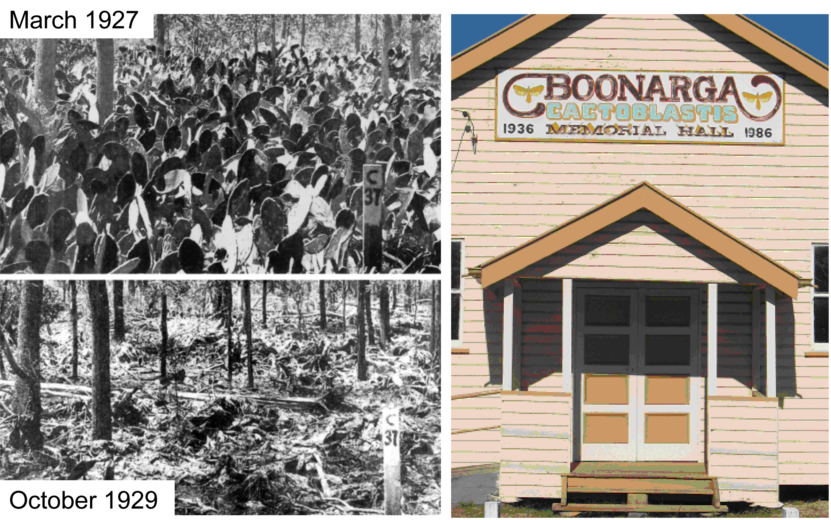
Chinnock RJ (2015) Feral opuntioid cacti in Australia. The State Herbarium of South Australia, Adelaide.

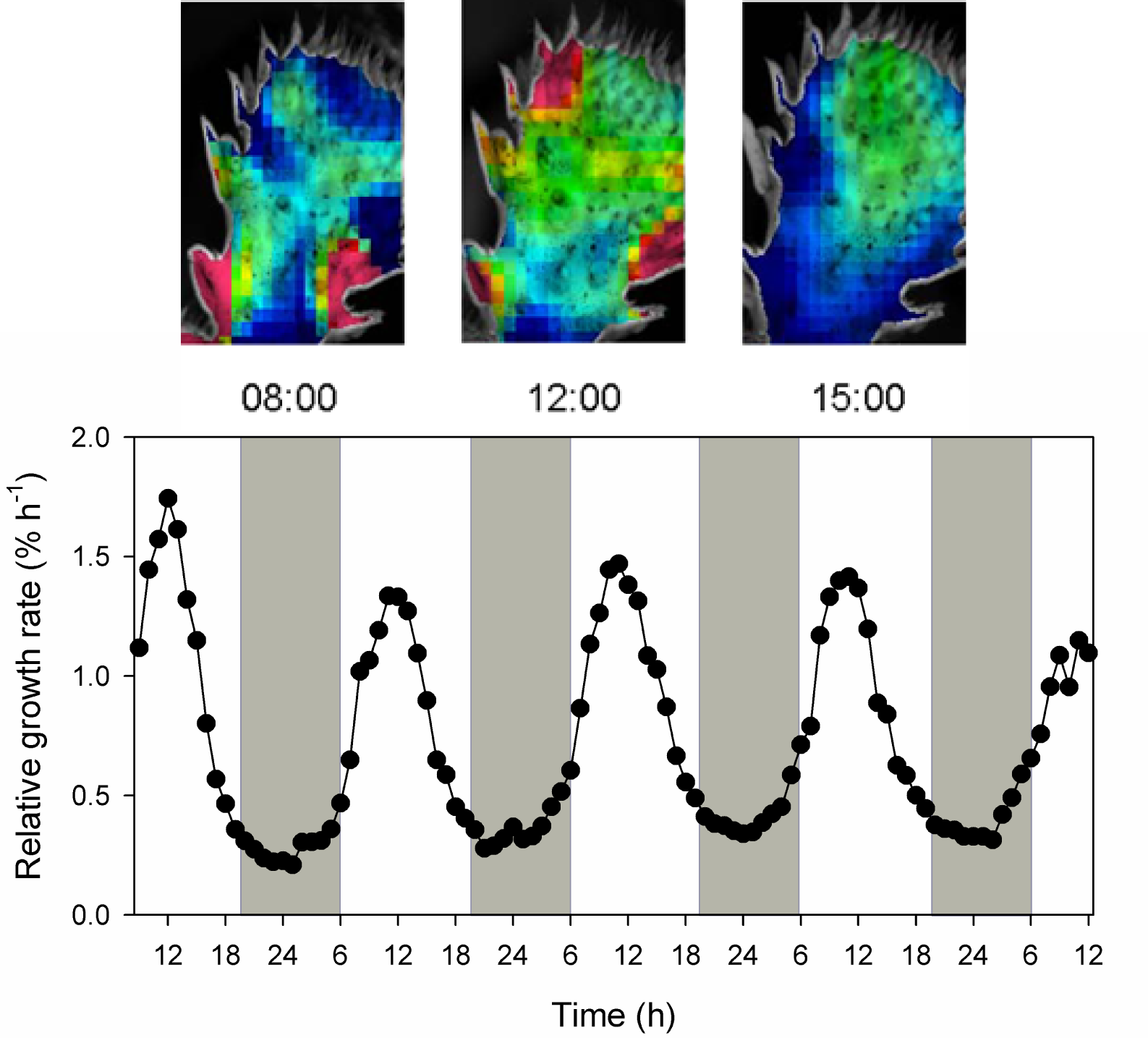
Gouws LM, Osmond CB, Schurr U et al. (2005) Distinctive diel growth cycles in leaves and cladodes of CAM plants: differences from C3 plants and putative interactions with substrate availability, turgor and cytoplasmic pH. Funct Plant Biol 32: 421-428

Griffiths H, Cousins AB, Badger MR et al. (2007) Discrimination in the dark: resolving the interplay between metabolic and physical constraints to phosphoenolpyruvate carboxylase during the crassulacean acid metabolism cycle. Plant Physiol 143: 1055-1067

Harvey A (2009) Draft state opuntioid cacti management plan. Government of South Australia, Adelaide.

Holtum JAM, Smith JAC, Neuhaus HE (2005) Intracellular transport and pathways of carbon flow in plants with crassulacean acid metabolism. Funct Plant Biol 32: 429-439

Keeley JE (2014) Aquatic CAM photosynthesis: a brief history of its discovery. Aquatic Bot 118: 38-44

Lüttge U (2002) CO2 concentrating: consequences in crassulacean acid metabolism. J Exp Bot 53: 2131-2142

Osmond CB, Neales T, Stange G (2008) Curiosity and context revisited: crassulacean acid metabolism in the Anthropocene. J Exp Bot 59: 1489-1502

Sage R (2014) Photosynthetic efficiency and carbon concentration in terrestrial plants: the C4 and CAM solutions. J Exp Bot 65: 3323-3325

von Caemmerer S, Griffiths H (2009) Stomatal responses to CO2 during a diel crassulacean acid metabolism cycle in Kalanchoe daigremontiana and Kalanchoe pinnata. Plant Cell Environ 32: 567-576

Walter A, Christ MM, Rascher U et al. (2008) Diel leaf growth cycles in Clusia spp. are related to changes between C3 photosynthesis and crassulacean acid metabolism during development and water stress. Plant Cell Environ 31: 484-491

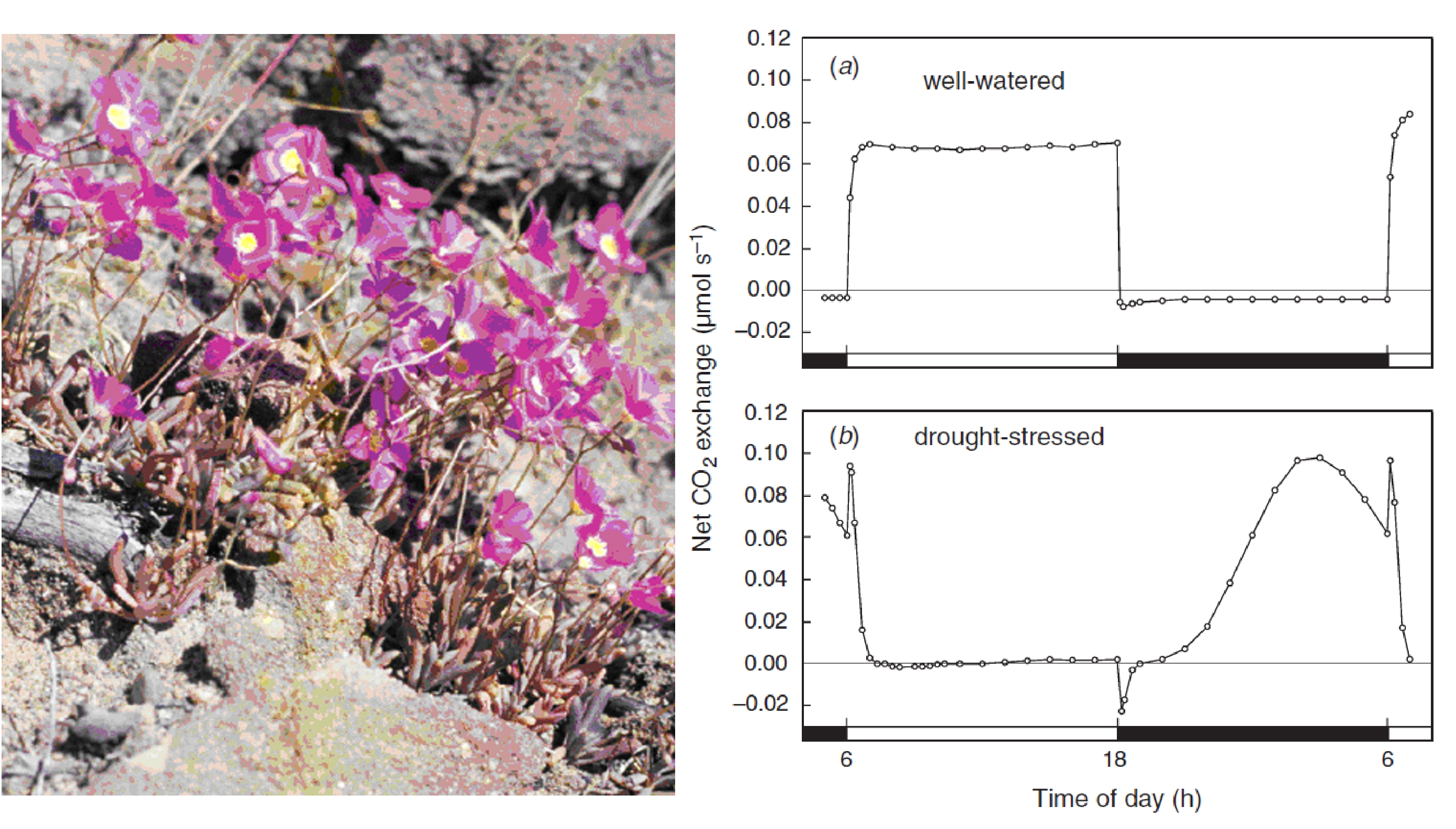
Winter K, Holtum JAM (2011) Induction and reversal of crassulacean acid metabolism in Calandrinia polyandra: effects of soil moisture and nutrients. Funct Plant Biol 38: 576-582

Winter K, Holtum JAM (2014) Facultative crassulacean metabolism (CAM) plants: powerful tools for unravelling the functional elements of CAM photosynthesis. J Exp Bot 65: 3425-3441

Winter K, Aranda J, Holtum JAM (2005) Carbon isotope composition and water-use efficiency in plants with crassulacean acid metabolism. Funct Plant Biol 32: 381-388

Winter K, Garcia M, Holtum JAM (2008) On the nature of facultative and constitutive CAM: environmental and developmental control of CAM expression during early growth of Clusia, Kalanchoë and Opuntia. J Exp Bot 59: 1829-1840

Winter K, Holtum JAM, Smith JAC (2015) Crassulacean acid metabolism: a continuous or discrete trait? New Phytol 208:  73-78

Yang X, Cushman JC, Borland AM et al. (2015) A roadmap for research on crassulacean acid metabolism (CAM) to enhance sustainable food and bioenergy production in a hotter, drier world. New Phytol 207: 491-504

 Zambrano VAB, Lawson T, Olomos E et al. (2014) Leaf anatomical traits which accommodate the facultative engagement of crassulacean metabolism in tropical trees of the genus Clusia. J Exp Bot 65: 3513-3523