Cell growth in plants occurs by division followed by expansion in special zones termed zones of division (meristems) or expansion or in trees, the cambium. “Growth” can be defined as the irreversible expansion of cell walls.
The process starts when cells divide in meristems and must expand before they can divide again, so expansion is often considered part of the division process. But the rapid part starts afterward in daughter cells from the division. If conditions are good, these cells can double in size every few hours and eventually become up to 100 or 1000 times larger than the original cells. This rapid enlargement is driven by osmotic forces as described below, but under strict developmental control.
In zones where expansion occurs, the cell wall is the controlling structure. It is tough and strong. Typically, osmosis creates turgor pressures (P) inside the cell, and the wall must be able to contain this pressure. Normally P is rather high, around 0.5 MPa or more or equivalent to two to three times the pressure in an automobile tyre. Cell wall yielding is a special property because it resists expansion sufficiently to maintain turgor pressure but yields sufficiently to allow cell expansion.
While the expansion occurs, new material is simultaneously synthesised and deposited in the wall. Maintaining wall thickness is essential because enlargement of 100- to 1000-fold would make the wall too thin to hold P. In most plant cells, the thickness remains the same within a factor of about two. Consequently, in the living cell, expansion and deposition go together in the wall. The cytoplasm also expands but most of the expansion is in the large central vacuole, and the cytoplasm ends up as a thin layer inside the wall. The plasma membrane and vacuole membrane expands, while the volume of the cytoplasmic organelles and their membranes most likely remains the same.
The direction of expansion is controlled by the alignment of cellulose microfibrils in transversely oriented bands (Figure 7.21). These permit extension to proceed only along the long axis of the cell. The cytoskeleton lying underneath the plasma membrane is a central player in co-ordination of wall shape and expansion in different dimensions (see Case Study 7.2 “The significance of cell walls”, Figure 3). The orientation of the arrays of microtubules determine the orientation of cellulose microfibrils in the wall.
7.3-Ch-Fig-7.21.jpg
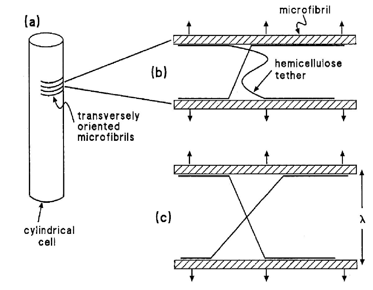
Figure 7.21 Schematic portrayal of the structure of an expanding cell wall. (a) A cylindrical cell, showing the transverse arrangement of cellulose microfibrils. (b) A pair of adjacent microfibrils connected by hemicellulose molecules (tethers) that are attached by hydrogen bonds; the arrows denote the force generated by turgor that is moving the microfibrils apart. One of the tethers, the straight one, is load-bearing; the other (wavy) is not. (c) The same microfibrils as in (b), whose separation in response to the applied force has resulted in a previously loose tether becoming load-bearing. (JB Passioura and SC Fry, Aust J Plant Physiol 19: 565-576, 1992)